


The cause of spontaneous abortion of normal conceptuses remains unknown in most embryos because of the difficulty of diagnosing too small embryos. We aimed to reveal latent liver abnormalities using novel phasecontrast radiographic computed tomography (PXCT). Embryos with liver volumes ≥ 2 SD above or below the mean for the stage of development were screened from 1156 MR images from the Kyoto Collection. Selected embryos were further analyzed by using PXCT. Liver abnormality was detected in 9 embryos by our protocol. Most of such liver abnormality embryos do not survive, as liver function becomes essential.
The cause of spontaneous abortion of normal conceptuses remains unknown in most embryos [1] because of the difficulty of obtaining appropriate embryo materials as well as diagnosing internal abnormalities in very small embryos. The crown-rump length of an embryo is ranging 23 to 32 mm during the embryonic period by Carnegie stage (CS) 23 (about 56–60 days after fertilization) [2], by which stage all primordia of the organs are already provided.
Many factors are involved in spontaneous abortion, not only the embryonic factor itself but also maternal health and the intrauterine environment affected by unspecified modulators. Such complicated factors distract attention from the laborious inspection of embryos, especially when embryos are externally normal.
More than 40,000 embryo and fetal specimens have been collected in the Kyoto Collection since 1961 for revealing the mechanism of congenital anomalies [3]. An MRI database of approximately 1200 well-preserved human embryos, diagnosed as externally normal, was acquired in 2000–2005 (the Kyoto embryo visualizing project) [4] to observe a number of normal human embryos. The database has proven to be useful for research purposes and as a teaching material [5].
Although the MRI database includes only externally normal embryo specimens, whether the internal organs were also normal cannot be guaranteed. Considering that the cumulative intrauterine mortality rate in normal conceptuses was estimated at 18% [1], the MRI database includes embryos that have potential abnormalities that would have led to spontaneous abortion. Thus, detailed observation of the internal organs of embryo specimens from the database may provide clues to spontaneous abortion in the embryonic and fetal periods. In this connection, we aimed to determine the latent abnormalities that may cause spontaneous abortion by using the MRI database and novel phase-contrast radiographic computed tomography (PXCT).
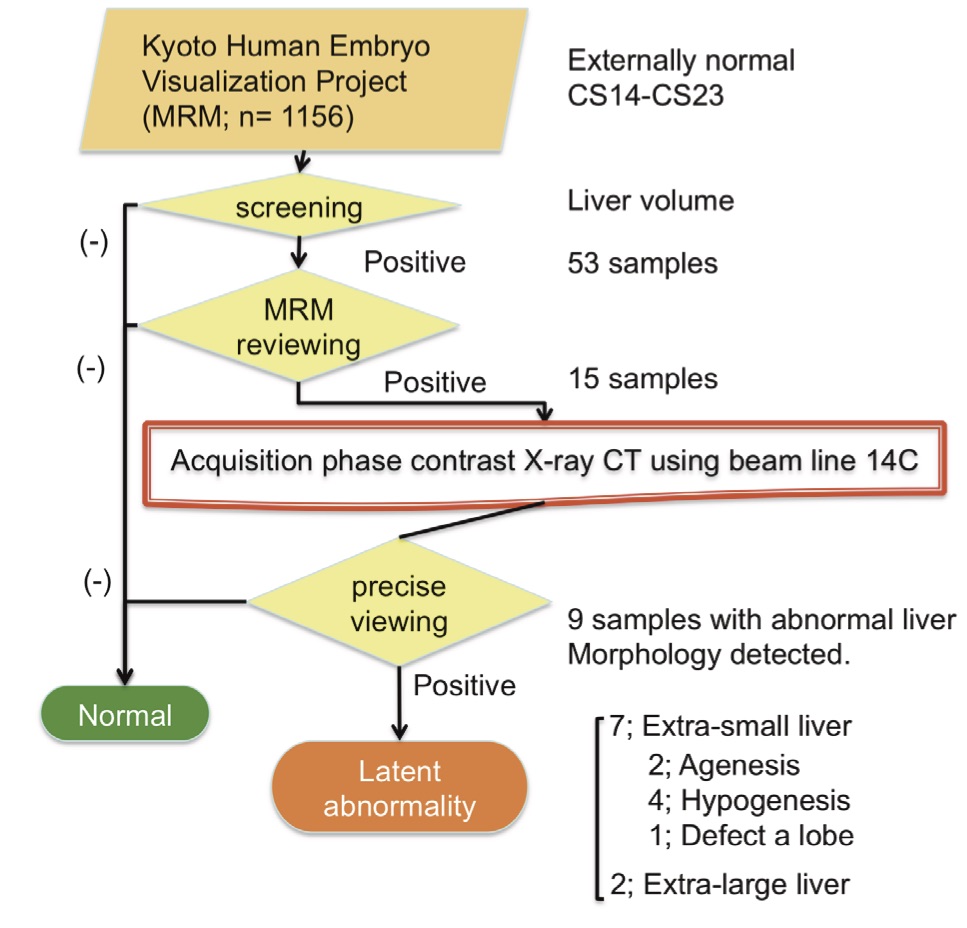
The MRI database was screened by using the volume of the liver as the target organ. Embryos with liver
volumes ≥ 2 SD above or below the mean for the stage of development were selected. Embryos with potentially abnormal livers were further analyzed by using PXCT. The PXCT data provide a resolution of ≥ 18 μm/pixel, which enabled highly sensitive measurement, approximately > 1000 times more sensitive than the conventional radiographic method using absorption contrast [6].
Liver abnormality was detected in 9 embryos after the procedure of our protocol (Fig. 1), which consisted of hepatic agenesis (2 embryos), hepatic hypogenesis (4), liver lobe defect (1), involvement of the liver to the thoracic cavity by diaphragm herniation (1), and other (1). Three embryos had only liver abnormalities and 6 exhibited complications in other organs. The prevalence of liver malformations in CS18 and CS21 in the intrauterine population of externally normal embryos is approximately 1.7%. Most of such liver abnormality embryos do not survive, as liver function becomes essential during development [7].
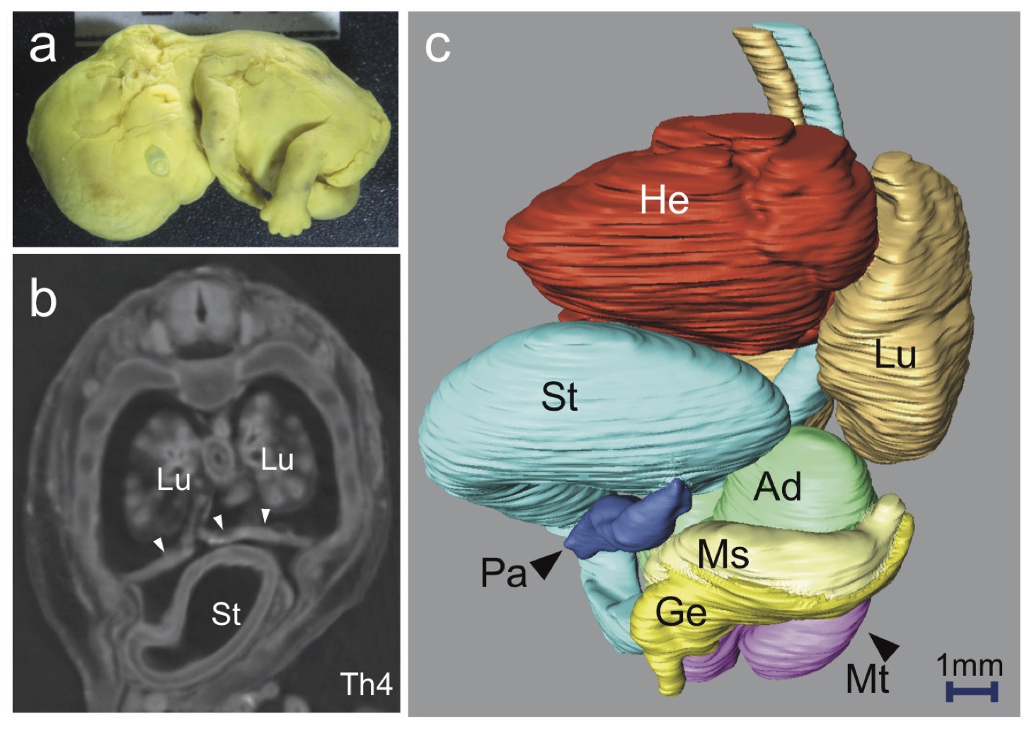
a External view of the embryo at CS21, which shows no obvious external abnormality. b Phase-contrast X-ray computed tomography (PXCT) transverse sections at the level of Th4. No liver (Li) was detected in the plane sections, while the stomach (St) was observed in the midsagittal transverse sections at Th4. The diaphragm is indicated by arrow heads. Lung (Lu). c Left anterior oblique view of the three-dimensional (3-D) PXCT reconstruction of the embryo using Amira software (Visage Imaging, Berlin, Germany), demonstrating the locations of all intrathoracic, retroperitoneal, and intra-abdominal organs.
The findings of note were as follows: agenesis of the liver; the stomach was deviated ventrally and cranially; the pancreas (Pa) was deviated ventrally; and the right mesonephros (Ms) and genital ridge (Ge) were absent. Adrenal gland (Ad), metanephros (Mt), and heart (He).
A representative embryo with liver agenesis is shown in Fig. 2. The size and gestational age were within the normal range for the embryo at CS 21. Obvious damage to, or anomalies of, the external forms were not present (Fig. 2a). The liver at CS21 usually occupies a large space in the abdominal cavity, which has a smooth surface due to the contact between the cranial surface and the diaphragm, and between the ventral surface and the abdominal wall [7]. In the present embryos, the liver was not detected in any of the serial plane sections (Fig. 2b). The locations of all intrathoracic, retroperitoneal, and intraabdominal organs were reconstructed in three dimensions (Fig. 2c). The absence of the liver had affected the locations of the other internal organs, especially the stomach, duodenum, and pancreas. The stomach was observed on the midsagittal line in the Th4 transverse sections, indicating that the stomach had deviated cranially and ventrally. The diaphragm was apparent in these sections.
The present study demonstrates that PXCT may be considered a powerful tool for visualization of internal structures of embryos, and for the detection of novel abnormalities during the embryonic period, without the need for histological analysis. The noninvasive and nondestructive properties of the technique are important for analysis of scarce specimens, such as human
embryos. The present study is the first step toward elucidating the latent abnormalities that result in spontaneous abortion in externally normal embryos [8].
REFERENCES
[1] K. Shiota, Congenital Anomalies 31, 67 (1991).[2] R. O’Rahilly and F. Müller, Developmental stages in human embryos: including a revision of Streeter’s” horizons” and a survey of the Carnegie Collection, (Carnegie Institution of Washington Publishing. 1987).
[3] H. Nishimura, K. Takano, T. Tanimura and M. Yasuda, Teratology 1, 281 (1968).
[4] K. Shiota, S. Yamada, T. Nakatsu-Komatsu, C. Uwabe, K. Kose, Y . Matsuda, T . Haishi, S. Mizuta and T . Matsuda, Am J Med Genet A 143A, 3121 (2007).
[5] T. Takakuwa, Antat Rec (2017), in press.
[6] A. Yoneyama, S. Yamada and T. Takeda, Fine Biomedical Imaging Using X-Ray Phase-Sensitive Technique, eds. G. D. Gargiulo and A. McEwan, Advanced Biomedical Engineering 107 (2011). [7] A. Hirose, T. Nakashima, S. Yamada, C. Uwabe, K. Kose and T. Takakuwa, Anat Rec 295, 51 (2012). [8] T. Kanahashi, S. Yamada, M. Tanaka, A. Hirose, C. Uwabe, K. Kose, A. Yoneyama, T. Takeda and T. Takakuwa, Anat Rec 299, 8 (2016).