 第127回日本解剖学会(2022.3.327-29)で発表しました。今年もon lineでした。
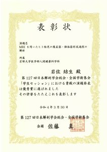
岩佐さんが学生セッション優秀賞に選ばれました。
ポスター討論
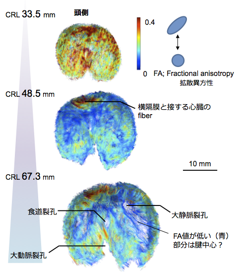
金橋 徹: 拡散テンソルイメージングを応用したヒト胚子期末から胎児期初期における横隔膜形成過程の解析
磯谷菜穂子: ヒト胚子期・胎児期初期の静脈管の形態観察と定量的解析
福井成美: ヒト胎児期初期における左心房形態形成~左心耳の形成と肺静脈が左心房に取り込まれる過程~
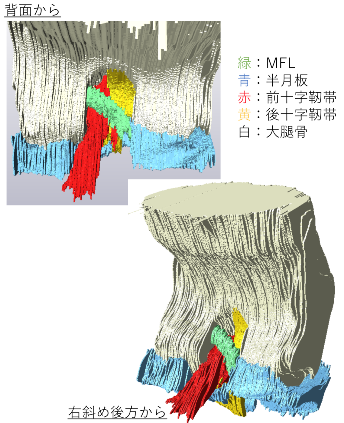
石田かのん:ラット胎仔の膝関節における半月大腿靭帯の形態形成
SAIZONOU Marie-Ange: Understanding of development and differentiation of the epithelium on Urinary Collecting System (UCS) (urothelium) in human embryonic metanephros; comparison with the urinary duct and bladder
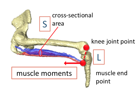
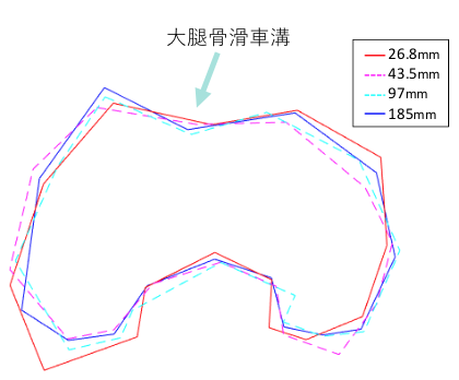
石川葵:ヒト胎児期における膝関節発生の三次元的解析
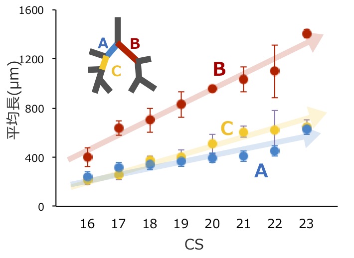
藤井瀬菜: ヒト胚子期における気管支樹の三次元的変化の定量的検討
学生セッション
岩佐結生: MRIを用いたヒト胎児の腹直筋・錐体筋形成過程の解析 *学生セッション優秀賞
中井尚一; ヒト胎児の主要血管径および血行動態の胎齢に伴う変化