


石田かのんさんの修士論文がCells Tissues Organsに掲載されました。
膝関節の後半月大腿靭帯(pMFL)は、膝関節安定への寄与や円盤状外側半月板(DLM)との関連が報告されていますが、健常な膝におけるその発生過程は調査されていません。本研究では、pMFLの形成について、ラットを対象に、組織切片、EFIC等を用いて検討しました。理学療法学講座の青山先生、谷間先生との共同研究です。
- EFIC画像から作成した3次元再構成画像を用いて、ラットの膝関節における後半月板靭帯の発達を解析し、他の膝関節構成要素との関係を検討。
- E16-21日のWistarラット胚の膝関節を対象。
- pMFLはE17から観察され、成熟ラットと同様にすべての発育段階で大腿骨内側顆と外側半月板に付着
- pMFLと膝関節周囲の構成要素は、発生初期から位置関係を維持したまま発達していると考えられる。
Tanima M, Ishida K, Ishikawa A, Yamada S, Takakuwa T, Aoyama T, Three-dimensional imaging analysis of the developmental process of posterior meniscofemoral ligaments in rat embryos. Cells Tissues Organs 2024, in press, , DOI: 10.1159/000536108
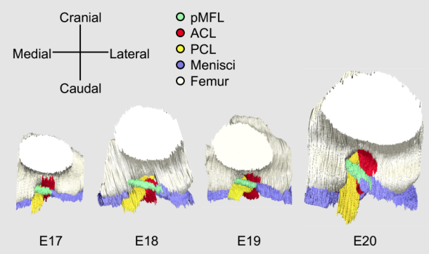
The posterior meniscofemoral ligament (pMFL) of knee joint is a ligament that runs posterior to the posterior cruciate ligament (PCL) and it is known that the height of the pMFL attachment site causes meniscus avulsion. Therefore, understanding the three-dimensional (3D) structure of the pMFL attachment site is essential to better understand the pathogenesis of meniscus disorders. However, the developmental process of pMFL has not been well investigated. The purpose of this study was to analyze pMFL development in rat knee jointsusing 3D reconstructed images produced from episcopic fluorescence image capture (EFIC) images and examine its relationship with other knee joint components. Knee joints of Wistar rat embryos between embryonic day (E) 16 and E21 were observed with HE stained tissues. Serial EFIC images of the hindlimbs of E17-E21 were respectively captured, from which 3Dimages were reconstructed and the features of pMFL structure: length and angle, were measured. Besides, the chronological volume changes and the volume ratio of the knee joint components compared to E17 were calculated to identify the differences in growth by components. pMFL was observed from E17 and was attached to the medial femoral condyle and lateral meniscus at all developmental stages, as in mature rats. The lack of marked variation in the attachment site and angle of the pMFL with the developmental stage indicates that the pMFL and surrounding knee joint components developed while maintaining their positional relationship from the onset of development. Current results may support to congenital etiology of meniscus disorder.