74. KanahashiT, ImaiH, Otani H, YamadaS, Männer J, Takakuwa T. Boundary Formation of the Human Caudal Foregut During the Early Fetal Period: Three-Dimensional Analysis Using T1-Weighted and Diffusion Tensor Images. Cells Tissues Organs, 2025, in press. doi: 10.1159/000546997
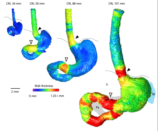
Introduction: While caudal foregut development in human fetuses has been outlined in previous research, the formation of its border region remains unclear. This study aimed to visualize the precise timeline of caudal foregut boundary formation. Methods: Three-dimensional images of the foregut from T1-weighted scans of 24 fetuses (crown–rump length [CRL]: 34–103 mm) were analyzed to measure the wall thickness and lumen diameter at nine specific sites. The internal structure in the border region was verified using histological sections and diffusion tensor imaging (DTI) tractography. Results: The lower esophageal and pyloric canal walls in samples with CRL ≥50 mm were relatively thicker. The esophageal wall at the esophageal hiatus, where the lower esophageal sphincter is located, was particularly thick in samples with CRL ≥88 mm. Increased wall thickness at the esophageal hiatus and pyloric canal resulted in a narrower lumen. The pyloric canal lumen narrowed from its distal to proximal sections. The lumen diameter-to-wall thickness ratio at the esophageal hiatus and proximal pyloric was negatively correlated with CRL. The thickened esophageal wall at the esophageal hiatus had a thick submucosa layer, and all layers in the pyloric canal thickened with growth. DTI tractography revealed that the lower esophageal wall mainly comprised longitudinal fibers, whereas the pyloric canal wall consisted solely of circular fibers, with fractional anisotropy increasing with growth. Conclusion: This study provides a comprehensive timeline of normal caudal foregut boundary formation during the early human fetal period, thereby improving the understanding of congenital foregut obstruction pathogenesis.
Ishida, N, Kanahashi T, Matsubayashi J, Imai, H, Männer J, Yamada S, Takakuwa,T. Change in diameters of the small intestine according to embryonic and early fetal growth. 2025. J Anatomy in press. doi: 10.1111/joa.14285.
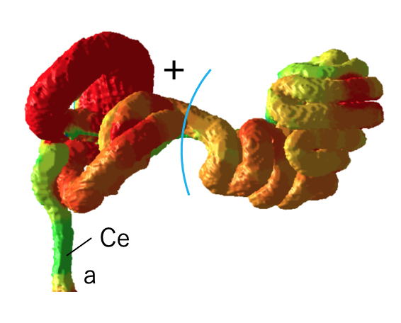
Abstract No previous studies have examined the diameter of the small intestine successively from the oral to the anal side of the small intestine. Therefore, the objectives of this study were to determine the successive intestinal diameters from the oral to the anal side (proximal to the distal) of the intestine, evaluate changes in diameter associated with growth, examine the effects of positional variation along the intestinal tract, investigate dynamic positional change from the extraembryonic coelom to the abdominal cavity, and assess the impact of complex tertiary intestinal loop formation. To this end, 14 human embryonic and fetal specimens with crown-rump lengths (CRLs) ranging from 25.6 to 69.0 mm were selected for high-resolution magnetic resonance imaging acquisition. The small intestines of the specimens were located in the extraembryonic coelom (herniation phase), transitioning phase, or abdominal cavity (return phase). The small intestine and mesentery were reconstructed in three dimensions, and the resulting morphological changes were observed and analyzed. Successive intestinal diameters from the oral to anal side of the small intestine were determined. Specifically, we observed the following: 1) gradual changes in the diameter of the position from the oral to the anal side in the jejunum-ileum, 2) the difference between the duodenum and jejunum-ileum, and 3) the difference between the superior part of the duodenum derived from the foregut and the remaining parts derived from the midgut. 4) Notably, the dynamic positional change from the extraembryonic coelom to the abdominal cavity, along with the rapid elongation and complex intestinal loop formation—a conspicuous phenomenon in the embryonic and early fetal periods—had little effect on the changes in diameter. This study indicates that increased diameter may serve as a useful indicator of intestinal development and differentiation, independent of tertiary intestinal loop formation and positional changes into and out of the abdominal cavity.
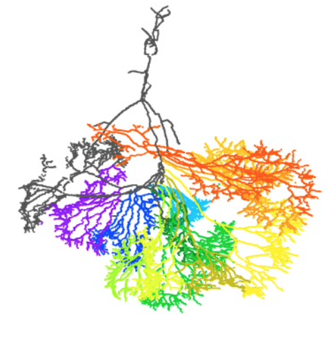
Introduction: Features of the superior mesenteric artery (SMA) and its intestinal branches during the embryonic and early fetal periods have not been fully described. We aimed to comprehensively elucidate the characteristics of intestinal branch artery formation in the SMA. Methods: Serial tissue sections of seven early fetal specimens belonging to the Blechschmidt Collection were digitalized and used for segmentation and reconstruction of the intestinal loop, SMA trunk, intestinal branch arteries, and mesentery for further analysis. Results: The intestinal branch arteries fed the intestinal tract from the oral side to the anal side, according to the order of their origin from the root to the periphery of the SMA trunk. SMA and intestinal branches were not as strongly conserved in their morphology as indicated in previous research but varied between specimens. Most intestinal branch arteries exhibited frequent branching with small intervals at the periphery, whereas the proximal branch exhibited few branches. Only a few peripheral branches made contact with the neighboring intestinal branch arteries. The fetal intestinal branch artery architecture differed greatly from that of adults. There were considerable inter- and intra-specimen variations in the intestinal tract length per feeding intestinal branch artery. The SMA branching arteries did not always supply each tertiary loop individually, and not every loop is connected to one branching artery. Conclusion: This study elucidates the characteristics of forming the SMA intestinal branch arteries. Specifically, the findings suggest that the SMA is similar to other arteries in that its branches show a level of variability in feeding tissues.
Takakuwa T,Kakeya M, Ishida N, Kanahashi T, Fujii S, Männer J, Yamada S. Superior mesenteric artery during intestinal loop formation and its positional changes from the extracoelom to the abdominal cavity. Cells Tissues Organs 2025, in press, DOI: 10.1159/000545751
■ 須藤紗帆、松林 潤、金橋 徹、今井 宏彦、大谷 浩、山田重人、高桑徹也;ヒト胚子期・胎児期初期における舌筋発生の検討 (The Development of the Tongue Muscles in the Human Embryonic and Fetal Period)
■ 八田 桃佳、金橋 徹、今井 宏彦、大谷 浩、山田 重人、高桑 徹也;拡散テンソル画像を用いた水晶体線維細胞の配向性の検討 (Analysis of lens fiber cells orientation in human embryonic and early fetal period using diffusion tensor imaging)
■ 石田 七彩、植田 優生、掛谷 真樹、松林 潤、金橋 徹、今井 宏彦、大谷 浩、山田 重人、高桑 徹也;中腸ループ形成を決定する要因:中腸の長さ、直径および位置の影響 (Factors determining midgut loop formation: The impact of midgut length, diameter, and location):
Graduate Student Presentation Award受賞しました!!
■ 熊谷 美優、金橋 徹、今井 宏彦、大谷 浩、多賀 厳太郎、高桑 徹也;高解像度MRIを用いたヒト胎児における大脳基底核原基の形成過程の検討 (Ganglionic Eminences Formation in the Human Fetus)
■ 青江 春菜、金橋 徹、今井 宏彦、大谷 浩、山田 重人、高桑 徹也;ヒト胎児期初期における上顎・下顎・歯胚の三次元解析 (Three-dimensional analysis of maxilla, mandible, and teeth at early human fetal stage)
■ 倭 友希、松田 幸樹、松林 潤、金橋 徹、今井 宏彦、米山 明男、山田 重人、高桑 徹也;ヒトの胚子期における足部・手部の形態形成 (Foot and hand morphogenesis during human embryonic development)
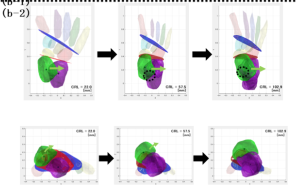
■ 金橋 徹、松林 潤、今井 宏彦、山田 重人、大谷 浩、高桑 徹也;ヒト胎児期初期における骨盤傾斜角の検討 (Analysis of human pelvic tilt angle in the early fetal period)
■ 藤井瀬菜、山田重人、高桑徹也:胚子の体軸がらせん状を描くか否か (Does the embryo’s body axis have a right-handed helical shape? : analysis of 3D reconstructions)
30. Three-dimensional structure of the human midgut with mesentery and factors determining midgut loop formation ヒト中腸と腸間膜の経時的構造変化および中腸ループ形成を決定する要因の検討 石田 七彩
29. Three-dimensional analysis of the human embryonic and early fetal lens 胚子期・胎児期初期におけるヒト水晶体の三次元解析 八田 桃佳
28. Development of the tongue in the human embryonic and fetal period 胚子期・胎児期初期におけるヒト舌の発生 須藤 紗帆
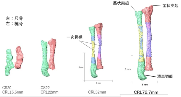
27. Three-dimensional analysis of human fetal tarsal development 胎児期におけるヒト足根骨の三次元解析 田村祥太郎
71. Ishida N, Ueda Y, Kanahashi T, Matsubayashi J, Imai H, Yamada S, Takakuwa T.Hierarchical loop formation in human midgut during physiological umbilical herniation, J Anatomy 2024, in press DOI:10.1111/joa.14228
CS16からCS18までのすべての標本で一次ループの形成が観察された。
腸間膜狭窄により中腸がを 4 つのセグメントに分割可能。
中腸の二次ループは、最初にCS19のセグメント2と4(S2とS4)で識別。
三次ループの形成は、CS21 で最初に確認
CS23 までに、三次ループはほとんどの標本のS2,3,4のセグメントで観察
S1は1つの二次ループのまま。
三次ループの数は頭殿長に応じて増加し、中腸の S2 から S4 で最大 19 個
ループ形成における遺伝的要因と生体力学的要因の役割を包括的に理解する上で極めて重要
Abstract
The primary loop, a single hairpin-shaped loop, becomes recognizable at the Carnegie stage (CS) 16. This loop projects toward the umbilical cord and subsequently gives rise to four secondary loops in the midgut of human embryos. As development advances, the segments corresponding to each secondary loop further develop into an increasing number of loops, referred to as tertiary loops. The mesenteric leaves and the narrowing parts, which serve as the borders of the secondary loops, remain identifiable throughout the subsequent stages of development. This study aimed to describe the morphological alterations that occur in the midgut and mesentery over time during the herniated phase of the midgut. A total of 47 human embryos between CS16 and CS23 and two fetuses in the physiological umbilical herniated stage were selected for high-resolution magnetic resonance imaging acquisition. Specimens were obtained from the Congenital Anomaly Research Center of Kyoto University. Serial tissue sections obtained from four embryos were subjected to histological observation. The midgut and mesentery were reconstructed in three dimensions, and the resulting morphological changes were observed and analyzed. Formation of the primary loop was observed in all specimens between CS16 and CS18. Secondary loops in the midgut were initially discerned at CS19 in segments 2 and 4 (S2 and S4). The border between S3 and S4 was identified at the apex of the midgut hernia, where traces of the vitelline artery and duct enter the mesentery. At CS21 and later stages of development, the presence of three borders at the exact location delineated by mesenteric narrowing was consistently observed, which resulted in the midgut being divided into four segments in all specimens. The formation of tertiary loops was initially identified at Carnegie stage (CS) 21, occurring in either segment S2 or S3. By CS23, tertiary loops were observed in three segments in most specimens. Notably, the initial formation of tertiary loops in S4 occurred one Carnegie stage later than in S2 or S3. Additionally, the increase in the number of folds and the length per fold in S4 was delayed compared with the number and length of folds observed in both S2 and S3. The number of loops in S1 remained constant (one secondary loop) across all specimens. Upon reaching a critical threshold length, the number of loops exhibited a marked increase, accompanied by rapid elongation in S2, S3, and S4. The number of tertiary loops increased in accordance with the crown-rump length, which exhibited a maximum of 19 tertiary loops in S2 to S4 of the midgut. These findings support the hypothesis that tertiary loops develop biomechanically through the rapid elongation of the midgut and slow growth of the mesentery. This study describes the morphological alterations occurring in the midgut and mesentery over time during the herniated phase of the midgut and provides a comprehensive understanding of the roles of genetic and biomechanical factors in loop formation.