|
|
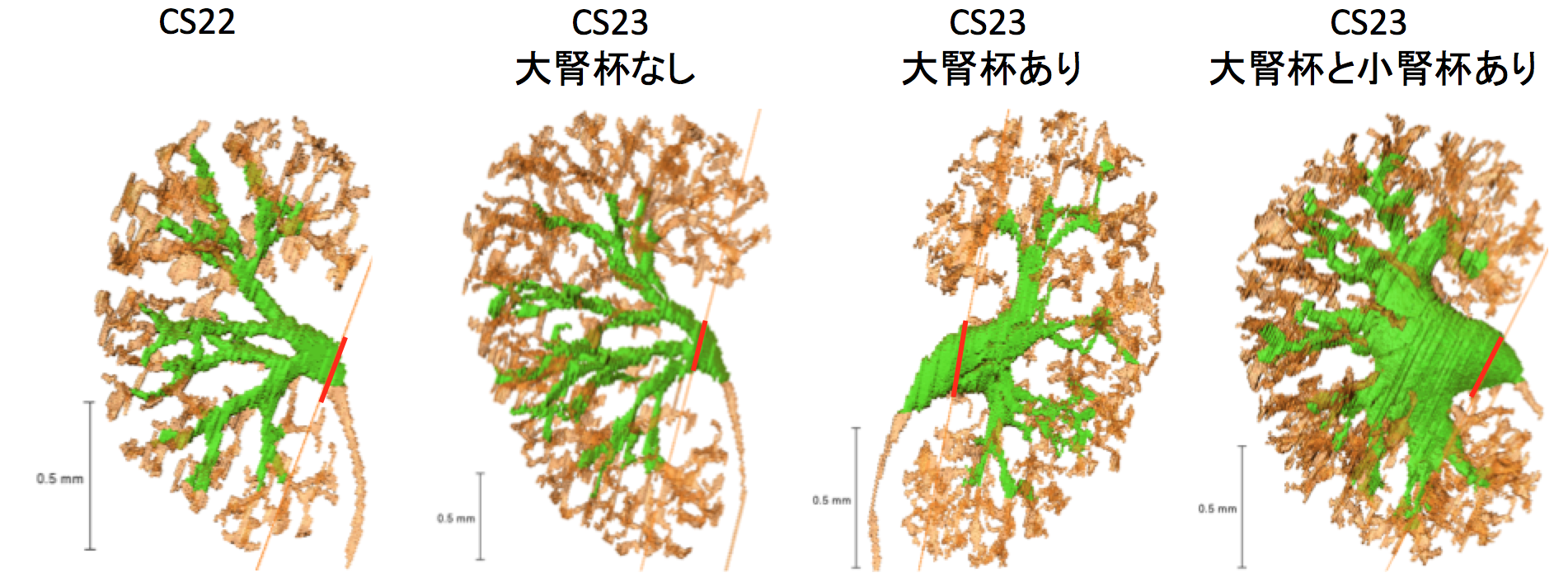
石山さんの修論がPLOS ONEに受諾、掲載されました。ヒト腎臓(後腎)における尿集合管系の形成過程を明らかにし実験動物(マウス)との差異を論じました。
- 最初の 尿路樹の分岐は CS16 で発生、CS23の最大分岐次数は12に達する、
- 二分分岐が急速に発生し、その後ネフロンの形成が続き、CS23において末端枝数あたりの糸球体数は1.34
- 腎盂拡張はCS23内でみられる
- 分岐次数は極部で高い
34. Ishiyama H, Ishikawa A, Kitazawa H, Fujii S, Matsubayashi J, Yamada S, Takakuwa T, Branching morphogenesis of the urinary collecting system in the human embryonic metanephros, PLoS ONE 13(9): e0203623. https://doi.org/10.1371/journal.pone.0203623
Abstract
An elaborate system of ducts collects urine from all nephrons, and this structure is known as the urinary collecting system (UCS). This study focused on how the UCS is formed during human embryogenesis. Fifty human embryos between the Carnegie stage (CS) 14 and CS23 were selected from the Kyoto Collection at the Congenital Anomaly Research Center of Kyoto University, Japan. Metanephroses, including the UCS, were segmented on serial digital virtual histological sections. Three-dimensional images were computationally reconstructed for morphological and quantitative analyses. A CS timeline was plotted. It consisted of the 3-D structural morphogenesis of UCS and quantification of the total amount of end-branching, average and maximum numbers of generations, deviation in the metanephros, differentiation of the urothelial epithelium in the renal pelvis, and timing of the rapid expansion of the renal pelvis. The first UCS branching generation occurred by CS16. The average branching generation reached a maximum of 8.74 ± 1.60 and was already the twelfth in CS23. The total end-branching number squared between the start and the end of the embryonic period. UCS would reach the fifteenth branching generation soon after CS23. The number of nephrons per UCS end-branch was low (0.21 ± 0.14 at CS19, 1.34 ± 0.49 at CS23), indicating that the bifid branching occurred rapidly and that the formation of nephrons followed after. The renal pelvis expanded mainly in CS23, which was earlier than that reported in a previous study. The number of nephrons connected to the UCS in the expanded group (246.0 ± 13.2) was significantly larger than that of the pre-expanded group (130.8 ± 80.1) (P < 0.05). The urothelial epithelium differentiated from the zeroth to the third generations at CS23. Differentiation may have continued up until the tenth generation to allow for renal pelvis expansion. The branching speed was not uniform. There were significantly more branching generations in the polar- than in the interpolar regions (P < 0.05). Branching speed reflects the growth orientation required to form the metanephros. Further study will be necessary to understand the renal pelvis expansion mechanism in CS23. Our CS-based timeline enabled us to map UCS formation and predict functional renal capacity after differentiation and growth.
Blechschmidt collectionスキャンプロジェクトについての論文がCongenital anomaliesに掲載されました。先天異常標本解析センター、ドイツ・ゲッティンゲン大学解剖学教室との共同プロジェクトです。
(ゲッティンゲン大学ヒト胚子コレクションの組織学的解析およびデジタルアトラス作成;基盤(B)(海外 )2015-2017)
- Blechschmidt コレクションの組織標本を、市販のフラットベッド スキャナーを使用して4800 dpiの解像度画像にデジタル化
- CRL64mm, CRL17.5mm (CS20) の2 つの標本について、最新の技術を使用して、立体再構成
⑦ Miyazaki R, Makishima H, Männer J, Sydow HG, Uwabe C, Takakuwa T, Viebahn C, Yamada S. The Blechschmidt Collection: revisiting specimens from a historical collection of serially sectioned human embryos and fetuses using modern imaging techniques, Congenit Anom, 2018, 58, 152-157, doi: 10.1111/cga.12261
ABSTRACT
Along with the Carnegie Collection in the United States and the Kyoto Collection in Japan, the Blechschmidt Collection (Georg-August-University of Göttingen, Germany) is a major historical human embryo and fetus collection. These collections are of enormous value to human embryology; however, due to the nature of the historical histological specimens, some stains are fading in color, and some glass slides are deteriorating over time. To protect these specimens against such degradation and ensure their future usefulness, we tried to apply modern image scanning and computational reconstruction. Samples of histological specimens of the Blechschmidt Collection were digitized into images using commercial flatbed scanners with a resolution of 4800 pixels per inch. Two specimens were reconstructed into three-dimensional (3D) images by using modern techniques to vertically stack two-dimensional images of the slices into 3D blocks. The larger specimen of crown-rump length (CRL) 64.0 mm, a series of very large histological sections in human embryology, was reconstructed clearly, with its central nervous system segmented before stacking. The smaller specimen of CRL 17.5 mm was also reconstructed into 3D images. The outer surface of the embryo was intact, and its development was classified according to the widely used Carnegie stages (CSs). The CS of the specimen was identified as the later half of CS 20. The invaluable Blechschmidt Collection can be revisited for further research with modern techniques such as digital image scanning and computational 3D reconstruction.
人間健康科学系専攻は、医療職の分野に貢献することが重要な使命の一つになっています。そのため、同分野で働いている方々が大学院に入学しやすいようないくつかの制度があります。
■ 修士課程
社会人特別選抜制度:入学時で3年以上(但し、看護科学コースの 志願者は5年以上)の志望分野に関連する医療実務経験を有する者については、申請時の手続きにより、試験の配点が変更できます。(2023年度入試より配点が変更になっています)
一 般 選 抜: 外国語(英語)100点、専門100点 、社会人特別選抜: 外国語(英語)100 点、専門 200 点
■ 博士課程
修士学位を取得していなくても、卒後の経歴(おもに研究活動)の内容によっては受験が可能になることがあります。(資格審査があります)
仕事をやめなくても博士課程に入学 できる可能性があります。
ご興味のある方は御連絡ください。
■医学研究科HP内の案内ページ>>(外部リンク)
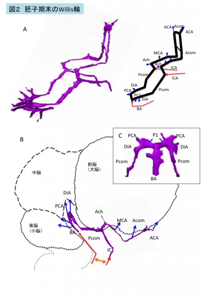
古市さんの卒論がAnat Recに掲載されました。
連続組織切片の 3-D 再構成を行い、胚子期末のWillis 輪( CW )の形成を検討しました。
- CW は胚子期末のすべての標本で閉鎖、
- CWは単一の平面でなく、複数の階段状の平面で構成
- 中脳と間脳の屈曲を反映してCW は尾部で急激に湾曲
- 85%(17/20)でvariation観察
- (anterior partのみ10例、anterior+posterior 6例、posteriorのみ1例)
- 観察されたvariationは、胎児、新生児、成人で報告されているものと同様
- CWのvariationが開始時期から存在することを示唆
33. Furuichi K, Ishikawa A, Uwabe C, Makishima H, Yamada S, Takakuwa T,
Variations of the circle of Willis at the end of the human embryonic period, 2018, 301, 1312-1319, doi:10.1002/ar.23794
ABSTRACT
Variations of the circle of Willis (CW) influence blood supply to the brain and adjacent structures in adults. We examined the formation of the CW in 20 human embryo samples at the end of the embryonic period using 3-D reconstructions of serial histological sections. The CW was closed in all samples, and did not form in a single plane, but was composed of multiple stair-like planes. The artery acutely curved at the caudal part of the CW, namely, at the inlet of the basilar artery and bifurcation of the P1 segment of the posterior cerebral artery (PCA), reflecting flexure of the mesencephalon and diencephalon at this stage. Variations were observed in 17 of 20 samples—only anterior parts (anterior communicating artery [Acom] and anterior cerebral artery [ACA]) in 10 samples, only posterior parts (posterior communicating artery [Pcom]) in one sample, and both anterior and posterior parts in six samples. Variations included the Acom formed as partially duplicated in three samples, duplicated in four, plexiform in three, and no channel as a result of a single azygos ACA in one. The ACA formed as duplicated in two, median ACA in two, and right hypoplasia in one. The Pcom formed in hypoplasia of either side in six samples. Variations observed in this study are similar to those observed in fetuses, neonates, and adults. The P1 segment of PCA was very large in all samples. The present observations indicate that variations in the CW are present from the initiation of CW formation.
 第58回日本先天異常学会で発表しました(7/27−7/29, 東京) 第58回日本先天異常学会で発表しました(7/27−7/29, 東京)
台風も近づき慌ただしい学会でした。
..
奨励賞受賞講演
・金橋 徹、山田 重人、田中 美玲、廣瀬あゆみ、上部千賀子、巨瀬 勝美、米山 明男、武田 徹、高桑 徹也
:大規模コレクションから潜在的な異常例を明らかにする新手法
 「多元計算解剖学」第4回サマーワークショップ(7月14、15日、神戸市)に参加しました。新学術領域研究「医用画像に基づく計算解剖学の多元化と高度知能化診断・治療への展開」研究班のmeetingです。今年度最終年になりました。 「多元計算解剖学」第4回サマーワークショップ(7月14、15日、神戸市)に参加しました。新学術領域研究「医用画像に基づく計算解剖学の多元化と高度知能化診断・治療への展開」研究班のmeetingです。今年度最終年になりました。
  佐賀県立九州シンクロトロン光研究センターに行きました。(2018.0612 佐賀県立九州シンクロトロン光研究センターに行きました。(2018.0612
位相CT撮像データからイメージ作成を行う手法を共同研究者の米山先生から習いました。
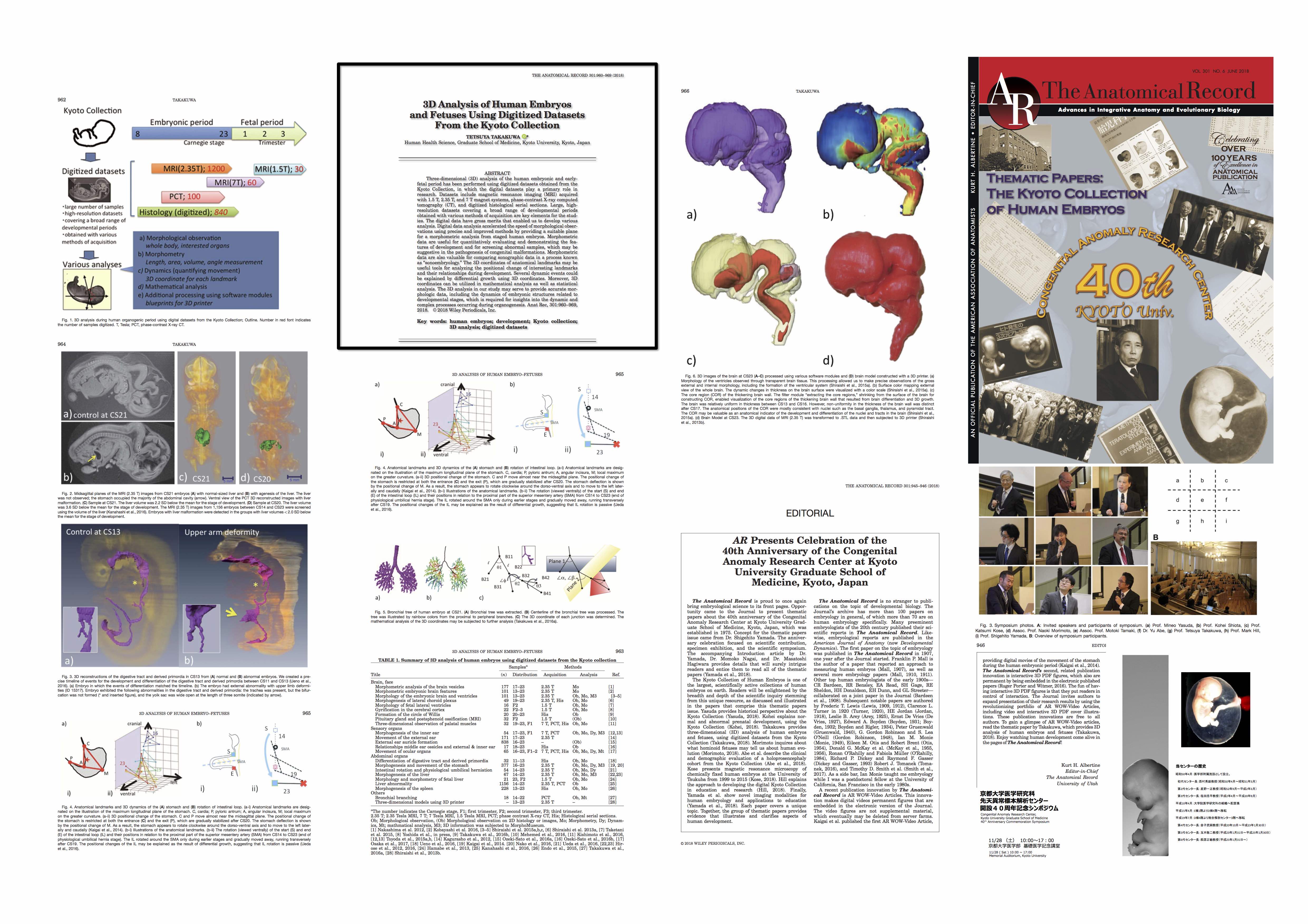
先天研開設40周年記念号がAnatomical Recに掲載されました。
先天研開設40周年記念号;ヒト胚子・胎児研究についての総説 Anat Rec 2018, 301,960-969 (クリックで拡大されます)
|
|