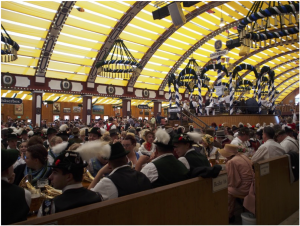
「多元計算解剖学」第3回サマーワークショップ(9月26、27日、神戸市)に参加しました。新学術領域研究「医用画像に基づく計算解剖学の多元化と高度知能化診断・治療への展開」研究班のmeetingです。
|
||||
  |
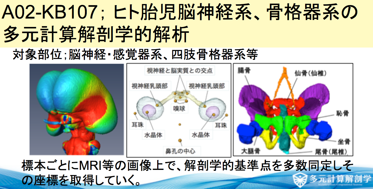
先天異常解析センター、ゲッチンゲン大学解剖学研究室との共同研究で、今年で3年目です。 今年度は9月の2-4週にのべ8名、本研究室からはM1の鈴木さん、西谷さんが参加しました。
130症例近くのガラス標本の撮像が終了しました。今後、成果をWeb等で公表する予定です。
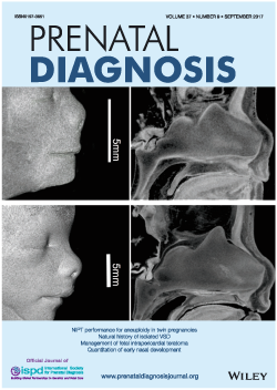 共同研究者の勝部先生の論文の図がprenatal Diagの表紙に採用されました。
⑭ Katsube M, Yamada S, Miyazaki R, Yamaguchi Y, Makishima H, Takakuwa T, Yamamoto A, Fujii Y, Morimoto N, Ito T, Imai H, Suzuki S, Quantitation of nasal development in the early prenatal period using geometric morphometrics and MRI: A new insight into the critical period of Binder phenotype. Prenatal Diag 37: 907–915, 2017, DOI: 10.1002/pd.5106.2017 AbstractObjectivesDisturbance of the development of the nasal septum in the early prenatal period causes congenital facial anomalies characterized by a flat nose and defects of the anterior nasal spine (ANS), such as Binder phenotype. The present research aimed to assess the development of the nasal septum and the ANS with growth in the early prenatal period. MethodsMagnetic resonance images were obtained from 56 specimens. Mid-sagittal images were analyzed by using geometric morphometrics for the development of the nasal septum, and angle analysis was performed for the development of the ANS. Additionally, we calculated and visualized the ontogenetic allometry of the nasal septum. ResultsOur results showed that the nasal septum changed shape in the anteroposterior direction in smaller specimens, while it maintained an almost isometric shape in larger specimens. Furthermore, mathematical evidence revealed that the maturation periods of the shapes of the ANS and the nasal septum were around 12 and 14 weeks of gestation, respectively. ConclusionThe anteroposterior development of the nasal septum is specific until 14 weeks of gestation, and it is important for nasal protrusion and the development of the ANS. Therefore, the disturbance of such development could induce low nasal deformity, including Binder phenotype. 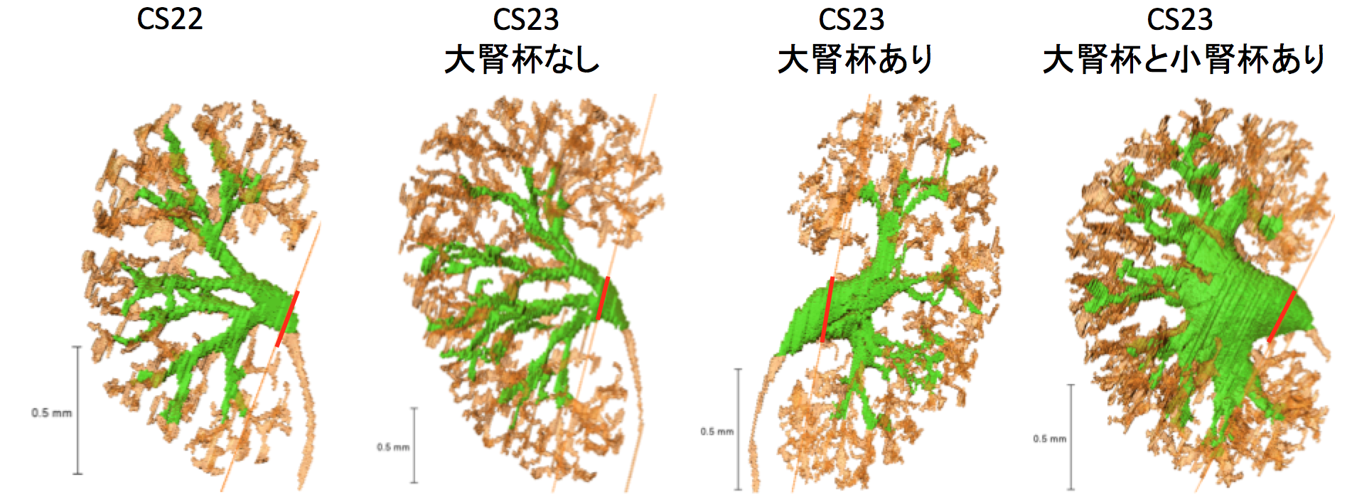 大腎杯はCS23に形成される 57回日本先天異常学会学術集会・第6回日本DOHaD学会 合同学術集会 ( 2017.8.26-28, 東京, 新宿)で発表しました。
学生部門で高石くんが日本病理学会総会最優秀賞を受賞しました。 英語口演
□ ヒト気管支発生の 3 次元的解析
Three-dimensional analysis of the bronchial branching in human embryonic stages
高桑徹也、村中太河、山田重人 他(急遽英語で発表しました!!)
ポスター
□ EFICを用いた膝関節の形態形成の解析(学生ポスター)日本病理学会総会最優秀賞受賞!!
Three-dimensional reconstruction of rat knee joint using episcopic fluorescence image capture
高石亮太、Xiangkai Zhang、青山朋樹、山田重人、高桑徹也
□ ヒト胚子期における脳形態形成の解析
Morphology and morphometry of the human embryonic brain: A three-dimensional analysis 白石直樹、片山愛里、中島崇、山田重人、上部千賀子、巨瀬勝美、高桑徹也
石山さん重責をご苦労様でした。 新学術領域研究「多元計算解剖学」第3回 国際シンポジウム(2017.3.8-9,奈良県文化会館) で発表しました。 A01-KB004 Three-dimensional Analysis of the Bronchial Branching in Human Embryonic Stages – Progress Overview FY2016 日本語
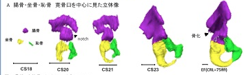 奥村さんの卒業研究がPLoS Oneに受諾されました。 ヒトの骨格形成は、保存しやすく、レントゲンでの解析が可能な骨化中、骨化後の解析がほとんどで、軟骨形成期の解析はほとんどされていません。今回、骨盤の軟骨形成期に着目し解析を進めました。
28. Okumura M, Ishikawa A, Aoyama T, Yamada S, Uwabe C, Imai H, Matsuda T, Yoneyama A, Takeda T, Takakuwa T, Cartilage Formation in the Pelvic Skeleton during the Embryonic and Early-Fetal Period, PLoS One 12(4): e0173852. https://doi.org/10.1371/journal. pone.0173852 [Open Access] Abstract The pelvic skeleton is formed via endochondral ossification. However, it is not known how the normal cartilage is formed before ossification occurs. Furthermore, the overall timeline of cartilage formation and the morphology of the cartilage in the pelvis are unclear. In this study, cartilage formation in the pelvic skeletons of 25 human fetuses (crown-rump length [CRL] = 11.9–75.0 mm) was observed using phase-contrast computed tomography and 7T magnetic resonance imaging. The chondrification center of the ilium, ischium, and pubis first appeared simultaneously at Carnegie stage (CS) 18, was located around the acetabulum, and grew radially in the later stage. The iliac crest formed at CS20 while the iliac body’s central part remained chondrified. The iliac body formed a discoid at CS22. The growth rate was greater in the ilium than in the sacrum-coccyx, pubis, and ischium. Connection and articulation formed in a limited period, while the sacroiliac joint formed at CS21. The articulation of the pubic symphysis, connection of the articular column in the sacrum, and Y-shape connection of the three parts of the hip bones to the acetabulum were observed at CS23; the connection of the ischium and pubic ramus was observed at the early-fetal stage. Furthermore, the degree of connection at the center of the sacrum varied among samples. Most of the pelvimetry data showed a high correlation with CRL. The transverse and antero-posterior lengths of the pelvic inlet of the lesser pelvis varied among samples (R2 = 0.11). The subpubic angle also varied (65–90°) and was not correlated with CRL (R2 = 0.22). Moreover, cartilaginous structure formation appeared to influence bone structure. This study provides valuable information regarding the morphogenesis of the pelvic structure. |
|||