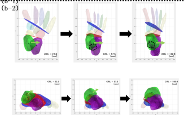
30. Three-dimensional structure of the human midgut with mesentery and factors determining midgut loop formation ヒト中腸と腸間膜の経時的構造変化および中腸ループ形成を決定する要因の検討 石田 七彩
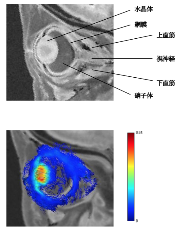
29. Three-dimensional analysis of the human embryonic and early fetal lens 胚子期・胎児期初期におけるヒト水晶体の三次元解析 八田 桃佳
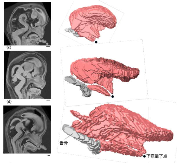
28. Development of the tongue in the human embryonic and fetal period 胚子期・胎児期初期におけるヒト舌の発生 須藤 紗帆
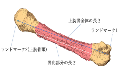
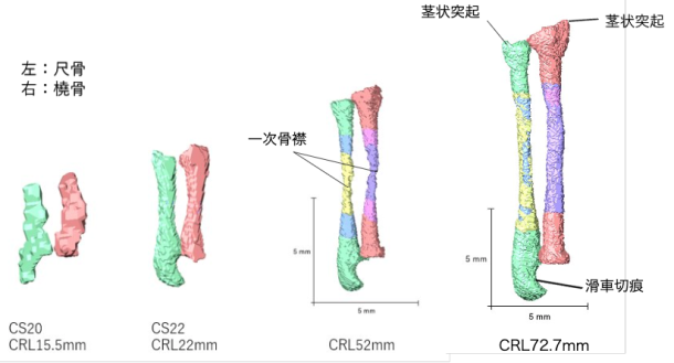
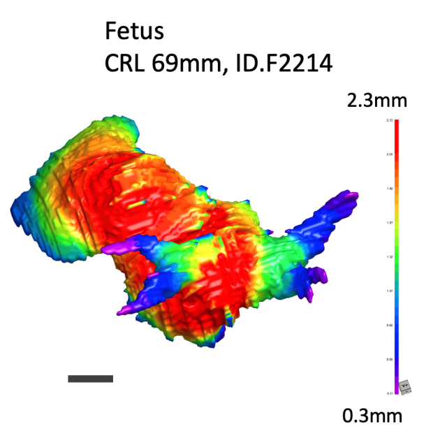
27. Three-dimensional analysis of human fetal tarsal development 胎児期におけるヒト足根骨の三次元解析 田村祥太郎
30. Three-dimensional structure of the human midgut with mesentery and factors determining midgut loop formation ヒト中腸と腸間膜の経時的構造変化および中腸ループ形成を決定する要因の検討 石田 七彩
藤井 瀬菜, 福井 成美, 金橋 徹, 松林 潤, 今井 宏彦, 米山 明男, 大谷 浩, 山田 重人, 高桑 徹也 ヒト胚子期から胎児初期における肺静脈と左心耳の形態形成 Morphogenesis of the pulmonary vein and left atrial appendage in human embryos and early fetuses 福井さんの修士研究を紹介いたしました。
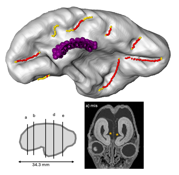
71. Kumagai M, Kanahashi M, Matsubayashi J, Imai H, Otani H, Takakuwa T. Primary sulci formation in human cerebral cortex development. Anat Rec (Hoboken) 2025, doi: 10.1002/ar.25637
Abstract
We aimed to determine the timing of appearance and the morphologic and morphometric features of the initial human cerebral sulcal formation. Using high-resolution magnetic resonance images obtained from 33 samples between 11 and 16 weeks (w) of gestation (crown-rump length <130 mm), the cerebral surface and internal structures on serial two-dimensional planes and all possible sulci on three-dimensional reconstructions were marked, allowing comparison of the positions of the sulci in the samples and inter-samples. Our method provided accurate conclusions regarding the timing of sulcal formation. Detection timing was as early as and earlier than those in previous studies using anatomical dissection and magnetic resonance imaging (MRI), respectively: <12 w for the callosum, <13 w for the hippocampal, calcarine, and parieto-occipital sulci, and <15 w for the lateral sulcus. Occasionally, an olfactory sulcus was detected. However, the cingulate sulcus could not be definitely identified. The lateral sulcus gradually appeared and changed shape. The lengths of the left and right sides of the olfactory sulci and the left side of the hippocampal sulcus increased linearly with the CRL. The length of the right side of the hippocampal sulcus and the left and right sides of the calcarine, parieto-occipital, and not determined_a sulci did not increase with the CRL The depth of the all sulci, except for the parieto-occipital sulci, increased linearly with the CRL. The sulci might not arise as if they elongate gradually but arise simultaneously over some distance. We determined the timing of the initial sulcal formation using high-resolution MRI. Our findings may significantly impact prenatal diagnosis and research on neurodevelopmental disorders.