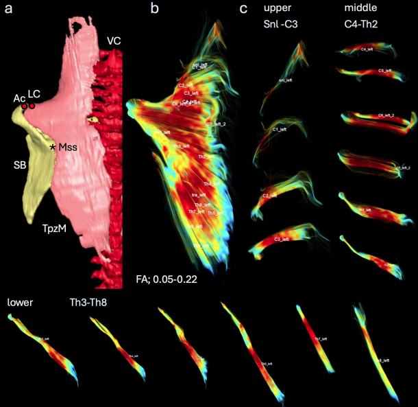
69. Iwasa Y, Kanahashi T, Imai H, Otani H, Yamada S, Takakuwa T. Human trapezius muscle development during early fetal period. J Anatomy 2024, 245, 663-673, doi: 10.1111/joa.14116
Abstract
J Anatomy 2024, 245巻, 11号(僧帽筋のDTI)
This study aimed to observe human trapezius muscle (TpzM) development during the early fetal period and apply diffusion tensor imaging (DTI) analysis to describe the muscle architecture that leads to physiological functions. Human embryonic and early fetal specimens were selected for this study. TpzM was first detected at Carnegie stage 20. The position of the TpzM changed with the formation of the scapula, clavicle, and vertebrae, which are its insertions and origins. DTI revealed the fiber orientation from each vertebral level to dissect each muscle. Fiber orientation in the ventral view gradually changed from the cervical to thoracic vertebrae, except for the middle part at which the insertions changed, which was almost similar in all early fetal specimens. The TpzM volume increased from C1 to C7 in the upper part, reached local maxima at C6 and C7 in the middle, and then decreased. These muscles can be categorized into three parts according to their insertions and presented with the features of each part. The fiber orientation and distribution of the three parts at the vertebral level were almost constant during the early fetal period. The border between the upper and middle parts was mainly located around the C6 and C7 vertebral levels, whereas the middle and lower parts were between the Th1 and Th2 vertebral levels. A three-dimensional change in the fiber orientation in the upper part of the TpzM according to the vertebral level was noticeable. Our data will help to elucidate the developmental processes of TpzM.
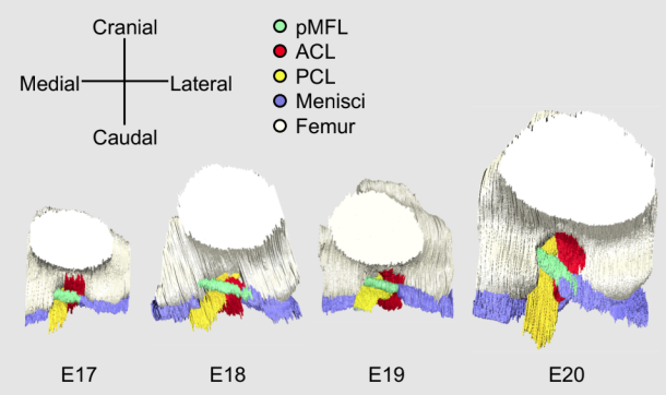
Tanima M, Ishida K, Ishikawa A, Yamada S, Takakuwa T, Aoyama T, Three-dimensional imaging analysis of the developmental process of posterior meniscofemoral ligaments in rat embryos. Cells Tissues Organs 2024, in press, , DOI: 10.1159/000536108
The posterior meniscofemoral ligament (pMFL) of knee joint is a ligament that runs posterior to the posterior cruciate ligament (PCL) and it is known that the height of the pMFL attachment site causes meniscus avulsion. Therefore, understanding the three-dimensional (3D) structure of the pMFL attachment site is essential to better understand the pathogenesis of meniscus disorders. However, the developmental process of pMFL has not been well investigated. The purpose of this study was to analyze pMFL development in rat knee jointsusing 3D reconstructed images produced from episcopic fluorescence image capture (EFIC) images and examine its relationship with other knee joint components. Knee joints of Wistar rat embryos between embryonic day (E) 16 and E21 were observed with HE stained tissues. Serial EFIC images of the hindlimbs of E17-E21 were respectively captured, from which 3Dimages were reconstructed and the features of pMFL structure: length and angle, were measured. Besides, the chronological volume changes and the volume ratio of the knee joint components compared to E17 were calculated to identify the differences in growth by components. pMFL was observed from E17 and was attached to the medial femoral condyle and lateral meniscus at all developmental stages, as in mature rats. The lack of marked variation in the attachment site and angle of the pMFL with the developmental stage indicates that the pMFL and surrounding knee joint components developed while maintaining their positional relationship from the onset of development. Current results may support to congenital etiology of meniscus disorder.
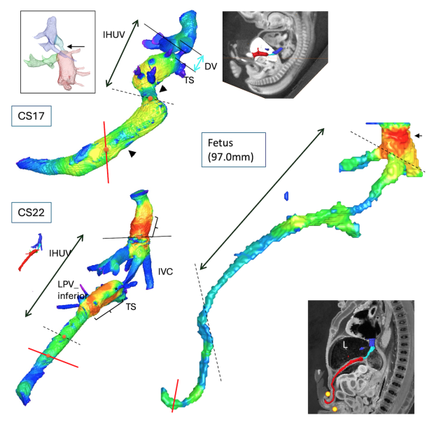
67. Isotani N, Kanahashi T, Imai H, Yoneyama A, Yamada S, Takakuwa T. Regional differences in the umbilical vein and ductus venosus at different stages of normal human development. Anat Rec (Hoboken), 2024, 307, 3306-3326.DOI:10.1002/ar.25421
During the fetal period, oxygenated blood from the placenta flows through the umbilical vein (UV), portal sinus, ductus venosus (DV), and inferior vena cava (IVC) to the heart. This venous route varies regionally in many aspects. Herein, we sought to characterize the venous route’s morphological features and regional differences during embryonic and early-fetal periods. Twenty-nine specimens were selected for high-resolution digitized imaging; 18 embryos were chosen for histological analysis. The venous route showed a primitive, large, S-shaped curved morphology with regional narrowing and dilation at Carnegie stage (CS) 15. Regional differences in vessel-wall differentiation became apparent from approximately CS20. The vessel wall was poorly developed in most DV parts; local vessel-wall thickness at the inlet was first detected at CS20. The lumen of the venous route changed from a non-uniform shape to a relatively round and uniform morphology after CS21. During the early-fetal period, two large bends were observed around the passage of the umbilical ring and at the inlet of the liver. The length ratio of the extrahepatic UV to the total venous route increased. The sectional area gradually increased during embryonic development, whereas differences in sectional area between the DV, UV, and IVC became more pronounced in the early-fetal period. Furthermore, differences in the sectional area between the narrowest part of the DV and other hepatic veins and the transverse sinus became more pronounced. In summary, the present study described morphological, morphometric, and histological changes in the venous route throughout embryonic and early-fetal development, clarifying regional characteristics.
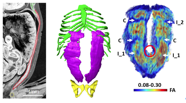
Iwasa Y, KanahashiT, ImaiH, OtaniH, YamadaS, Takakuwa T. Formation of tendinous intersections in the human fetal rectus abdominis, J Anatomy 2024, in press, DOI: 10.1111/joa.14064
Abstract
Previous studies have poorly described the initial development process of the tendinous intersections of the rectus abdominis muscle (RAM). The present study aimed to observe the formation of tendinous intersections in the RAM during the early fetal period using diffusion tensor imaging (DTI). Fifteen human fetal specimens (crown-rump length [CRL]: 39.5–93.7 mm) were selected. Three-dimensional measurements revealed that Zone-4 (i.e., the zone between the pubic symphysis and the caudal base of the umbilical ring in the RAM) had a smaller width and was thicker than Zone-1 and Zone-2 (i.e., the zones between the costal arch and the cranial base of the umbilical ring) and Zone-3 (i.e., the zone at the umbilical ring). Characteristics of tendinous intersections in the RAM during the early fetal period were assessed according to number, size, type, laterality, and sex. The mean number of tendinous intersections on both sides was 3.1 (range: 2.0–4.0), and 21% of specimens had only two tendinous intersections, which was higher than that reported in previous adult studies. The present data suggest that the formation of tendinous intersections was still in progress in specimens with two tendinous intersections in the RAM and that the third tendinous intersection was formed in Zone-2. Ordinal logistic regression via generalized estimating equations revealed that the odds for a higher type of tendinous intersections in Zone-1 and Zone-2 were significantly higher than those in Zone-4 (adjusted odds ratio: 14.85, 8.84). The odds for the presence of incomplete types (tendinous intersections that could not completely transverse the RAM) in Zone-3 were significantly higher than those in Zone-1 (adjusted odds ratio: 7.4). The odds for missing tendinous intersections in Zone-4 were significantly higher than those in Zone-1 (adjusted odds ratio: 20.5). These zonal differences in the formation of tendinous intersections were consistent with those observed in previous adult studies. In this study, DTI detected tendinous intersections in a sample with a CRL of 45.8 mm (approximately 11 weeks of gestation), which is earlier than that in previous histological findings, indicating that the RAM does not have mature tendinous intersections until the 17th week of gestation. In conclusion, DTI could detect the premature differentiation of tendinous intersection formation. Our data may aid in elucidating the developmental processes of tendinous intersections in the RAM.
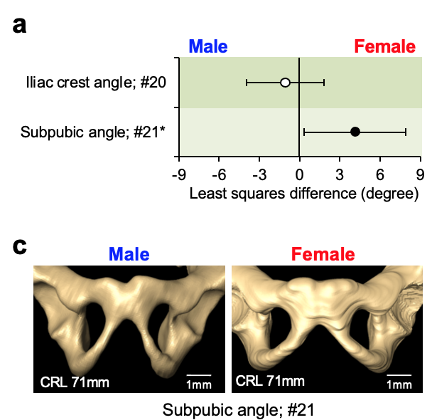
Kanahashi T, Matsubayashi J, Imai H, Yamada S, Otani H, Takakuwa T. Sexual dimorphism of the human fetal pelvis exists at the onset of primary ossification, Communications Biology, 2024, 7:538, https://doi.org/10.1038/s42003-024-06156-y
Abstract Human adolescent and adult skeletons exhibit sexual dimorphism in the pelvis. However, the degree of sexual dimorphism of the human pelvis during prenatal development remains unclear. Here, we performed high-resolution magnetic resonance imaging-assisted pelvimetry on 72 human fetuses (males [M]: females [F], 34:38; 21 sites) with crown-rump lengths (CRL) of 50–225 mm (the onset of primary ossification). We used multiple regression analysis to examine sexual dimorphism with CRL as a covariate. Females exhibit significantly smaller pelvic inlet anteroposterior diameters (least squares mean, [F] 8.4 mm vs. [M] 8.8 mm, P = 0.036), larger subpubic angle ([F] 68.1° vs. [M] 64.0°, P = 0.034), and larger distance between the ischial spines relative to the transverse diameters of the greater pelvis than males. Furthermore, the sacral measurements indicate significant sex-CRL interactions. Our study suggests that sexual dimorphism of the human fetal pelvis is already apparent at the onset of primary ossification.