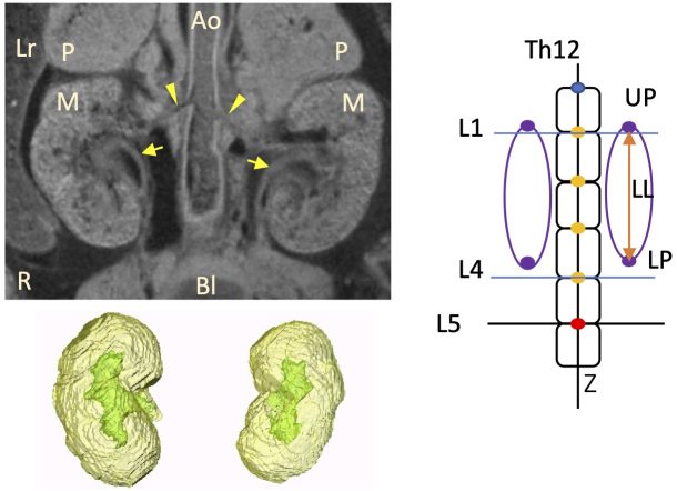
Ishiyama-Takara H, Matsubayashi J, Yamada S, Tetsuya Takakuwa T, Height difference between the right and left metanephroi during early human fetal development, Congenit Anom 64(3) 164-166, 2024.
Saizonou MA, Kitazawa H, Kanahashi T, Yamada S, Takakuwa T, Epithelial development of the urinary collecting system in the human embryo, PLOS ONE 19(4): e0301778. https://doi.org/10.1371/journal.pone.0301778
Abstract
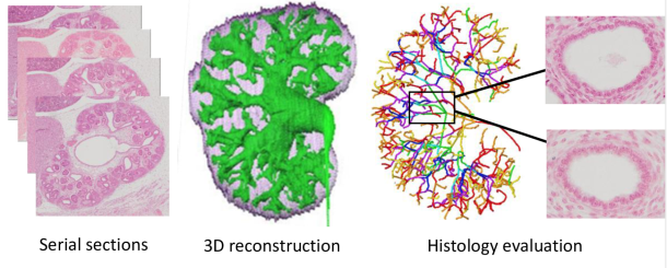
The urinary collecting system (UCS) consists of organized ducts that collect urine from the nephrons and transport it to the ureter and bladder. Understanding the histogenesis of the UCS is critical. Thirty human embryos between the Carnegie stages (CS) 18 and 23 were selected from the Congenital Anomaly Research Center, Kyoto, Japan. Epithelia of the UCS, ureter, and bladder of each sample were randomly selected. Histological findings of the epithelia were analyzed according to the following criteria: type of epithelium, presence or absence of glycogen, percentage of migrated nuclei, percentage of cells in mitosis, and the surrounding mesenchyme. A thickened epithelium lining a narrow luminal cavity was observed in the pre-expanded pelvic specimens at CS18-CS23. At CS23, after pelvic expansion, the UCS showed a thin epithelium with a large luminal cavity mainly located on the early branches, whereas the epithelium covering the subsequent branches had medium thickness. Histological characteristics differed depending on the UCS part and sample stage. The degree of differentiation was evaluated, revealing that in CS18-CS23 pre-expanded pelvis specimens, the undifferentiated epithelium was found in the zeroth to third/fifth generation, whereas at CS23, after pelvic expansion, a differentiated epithelium covered the UCS zeroth to seventh generation. In a comparison of the urothelial epithelium between the UCS, ureter, and bladder, we found that urinary tract differentiation may be initiated in the bladder, followed by the ureter, UCS zeroth to seventh generations, and finally, UCS eighth to end generations. An understanding of the histogenesis of embryonic stage UCS can aid in the clinical management of congenital urinary tract defects and other diseases.
Marie Ange Saizonou, Haruka Kitazawa, Toru Kanahashi, Shigehito Yamada, Tetsuya Takakuwa; Epithelial development of the urinary collecting system in the human embryo
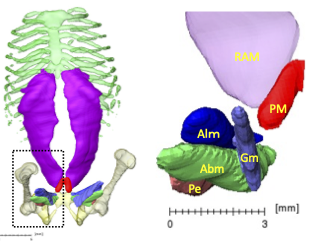
The pyramidalis muscle (PM) is a paired small triangular muscle of the anterior abdominal wall, the physiological significance of which remains unclear. Recent studies have failed to detect this muscle during the embryonic period. Hence, the present study aimed to determine when PM emerged and reveal its features using high-resolution magnetic resonance imaging. Fourteen embryos between Carnegie stage (CS)18 and CS23 and 59 fetuses (crown-rump length: 39.5–185.0 mm) were selected for this study. The PM was first detected in one of the three samples at CS20. It was detected in five of the seven samples (71.4%) between CS21 and CS23. Forty-eight samples (81.4%) at early fetal period had PMs on both the right and left sides, and three (5.1%) had that only on the right side. Eight samples (13.6%) had no PMs. No side-differences or sexual dimorphisms were detected. The PM length was larger than the width in most samples, although the length/width ratio varied among the samples. The PM/rectus abdominis muscle length and PM/umbilicus-pubic symphysis length ratios were almost constant, irrespective of the crown-rump length. The PM is located ventrally inferior to the rectus abdominis and closer to the medial muscle groups of the lower limb than the rectus abdominis. The present study demonstrated that PM formation occurred in the late embryonic period, and that the frequency, side differences, sex dimorphism, and spatial position of the PM in the early fetal period were similar to those in adults.