2017年度;卒業研究発表会が行われました(杉浦ホール)。
ヒト胚子期における後頸部の膨隆の組織学的検討 大賀 彩子
ヒト胚子期における肋骨と胸郭の三次元的解析 奥野 香澄 (石津研)
後腎における腎小体形成の組織学的検討 北沢 遥
ヒト胚子期~胎児期初期における肩甲骨の形態形成 坂本 梨乃
生理的臍帯ヘルニアの還納過程及び還納後の小腸の位置固定 長田 明香里
|
||||
  |
2017年度;卒業研究発表会が行われました(杉浦ホール)。
ヒト胚子期における後頸部の膨隆の組織学的検討 大賀 彩子 ヒト胚子期における肋骨と胸郭の三次元的解析 奥野 香澄 (石津研) 後腎における腎小体形成の組織学的検討 北沢 遥 ヒト胚子期~胎児期初期における肩甲骨の形態形成 坂本 梨乃 生理的臍帯ヘルニアの還納過程及び還納後の小腸の位置固定 長田 明香里
白石直樹くんの博士審査会が行われました。 (11月27日18時 高井ホール於) Morphology and morphometry of the human embryonic brain: A three-dimensional analysis (ヒト胚子期における脳の三次元形態計測学的解析) (NeuroImage, 2015, 115, 96-103、掲載済み) ヒトの発生時期のうち、組織と器官を急速に形成する器官形成期は先天異常の発生の可能性が高い時期であり解析に重要な時期である。京都大学大学院医学研究科附属先天異常標本解析センターが保有する外見上、傷や先天異常がないヒト胚子群から、2.35TのMR顕微鏡で撮像されたCarnegie Stage(CS)13~23のヒト胚子立体画像を用いて、脳実質と脳室の形態形成について立体化像の形態変化の詳細な観察と定量学的な検討を加えた。脳実質の厚みの変化を色彩表示する方法、及び内外から大脳壁の厚さ分を削り、残った部分を肥厚部として描出するという方法を用いて可視化した。脳実質の体積はCS13からCS23間に164.4倍に形態変化を伴いながら著しく増大した。前脳、中脳、菱脳の体積もそれぞれ増大した。小脳はCS20に初めて観察され、菱脳に対する小脳の体積比はCS20の約7.2%から、CS23では12.8%と大きく増大していた。胚子体積に対する脳実質体積比はCS15からCS23の期間で11.6〜15.5%と大きな変化はなかった。前脳部の肥厚部はCS16から見られ、その解剖学的位置は大脳基底核、視床、内包と合致した。CS17以降には神経核が発達し、特に前脳部の大脳基底核付近、菱脳部の基板や小脳部が不均一に肥厚していくことがわかった。CS20になると前脳部や菱脳部で厚さの不均一性が目立ち、CS23では著明であった。これらの不均一な肥厚が、胚子期における脳の複雑な屈折や進展に影響を及ぼしている可能性がある。今回、提示した手法は神経核の発達を観察する上で非常に優れた方法であり、胎児期の脳発達の観察、解析に応用可能と考えられる。以上の研究は器官形成期のヒト脳の発生の解明に貢献し人体発生学、脳発生学や胎児診断に寄与するところが多い。
大坂さんの修論の前半部がAnatomical Recordに掲載されました。 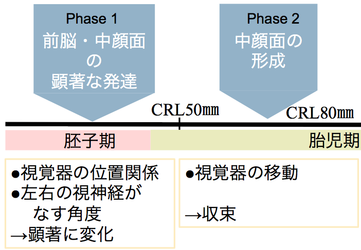 頭部・顔面形成にともなう眼の位置変化を胚子期から胎児期初期にかけて解析しました。
Osaka M, Ishikawa A, Yamada S, Uwabe C, Imai H, Matsuda T, Yoneyama A, Takeda T, Takakuwa T, Positional changes of the ocular organs during craniofacial development, Anatomical Record, 300(12), 2107–2114, 2017 DOI: 10.1002/ar.23588 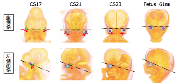 ABSTRACTThe present study aimed to describe the positional changes of the ocular organs during craniofacial development; moreover, we examined the relationships among the ocular organs and other internal structures. To do this, we traced the positions of the ocular organs in 56 human early fetal samples at different stages of development using high-resolution magnetic resonance imaging and phase-contrast X-ray computed tomography. The eyes were located on the lateral side in the ventral view at Carnegie stage (CS) 16, and then changed their positions medially during development. The eyes remained in the neurocranium until CS17. However, the eyes changed their positions medially and caudally in the viscerocranium after CS18. The positional relationship between the eyes and pituitary gland changed in the lateral view as development progressed. Specifically, they were close to each other at CS17, but moved apart during the later stages of development. These positional changes were also demonstrated quantitatively with morphometric analyses. Based on the present data, the positional changes of the eyes can be categorized into phases, as follows: Phase 1, dramatic positional changes (early fetal period until CS23); and Phase 2, mild positional changes (stabilized; early fetal period after CS23). Notably, all absolute lengths measured in the present study linearly increased as the crown-rump length increased irrespective of the phase, while features of the measured angles and ratios differentially changed in Phases 1 and 2. The present data may help improve our understanding of both the normal and abnormal development of the ocular organs and craniofacial area. 「多元計算解剖学」第3回サマーワークショップ(9月26、27日、神戸市)に参加しました。新学術領域研究「医用画像に基づく計算解剖学の多元化と高度知能化診断・治療への展開」研究班のmeetingです。
先天異常解析センター、ゲッチンゲン大学解剖学研究室との共同研究で、今年で3年目です。 今年度は9月の2-4週にのべ8名、本研究室からはM1の鈴木さん、西谷さんが参加しました。
130症例近くのガラス標本の撮像が終了しました。今後、成果をWeb等で公表する予定です。
 共同研究者の勝部先生の論文の図がprenatal Diagの表紙に採用されました。
⑭ Katsube M, Yamada S, Miyazaki R, Yamaguchi Y, Makishima H, Takakuwa T, Yamamoto A, Fujii Y, Morimoto N, Ito T, Imai H, Suzuki S, Quantitation of nasal development in the early prenatal period using geometric morphometrics and MRI: A new insight into the critical period of Binder phenotype. Prenatal Diag 37: 907–915, 2017, DOI: 10.1002/pd.5106.2017 AbstractObjectivesDisturbance of the development of the nasal septum in the early prenatal period causes congenital facial anomalies characterized by a flat nose and defects of the anterior nasal spine (ANS), such as Binder phenotype. The present research aimed to assess the development of the nasal septum and the ANS with growth in the early prenatal period. MethodsMagnetic resonance images were obtained from 56 specimens. Mid-sagittal images were analyzed by using geometric morphometrics for the development of the nasal septum, and angle analysis was performed for the development of the ANS. Additionally, we calculated and visualized the ontogenetic allometry of the nasal septum. ResultsOur results showed that the nasal septum changed shape in the anteroposterior direction in smaller specimens, while it maintained an almost isometric shape in larger specimens. Furthermore, mathematical evidence revealed that the maturation periods of the shapes of the ANS and the nasal septum were around 12 and 14 weeks of gestation, respectively. ConclusionThe anteroposterior development of the nasal septum is specific until 14 weeks of gestation, and it is important for nasal protrusion and the development of the ANS. Therefore, the disturbance of such development could induce low nasal deformity, including Binder phenotype. 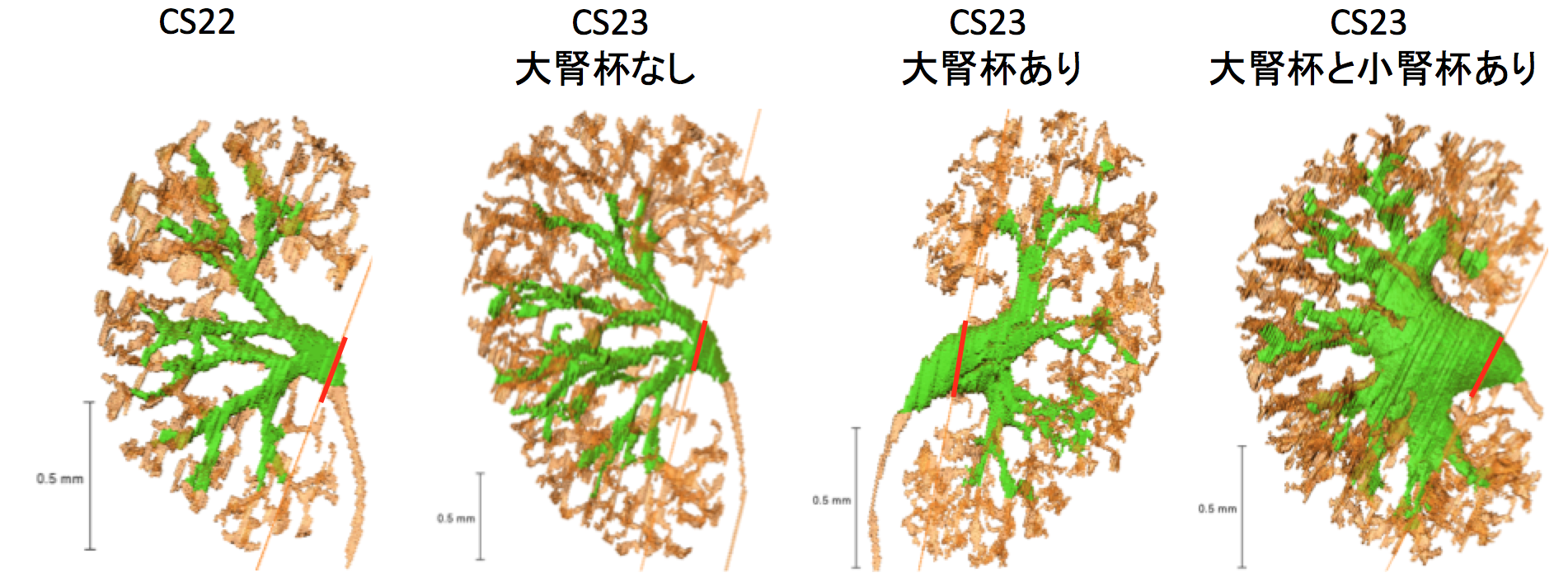 大腎杯はCS23に形成される 57回日本先天異常学会学術集会・第6回日本DOHaD学会 合同学術集会 ( 2017.8.26-28, 東京, 新宿)で発表しました。
学生部門で高石くんが日本病理学会総会最優秀賞を受賞しました。 英語口演
□ ヒト気管支発生の 3 次元的解析
Three-dimensional analysis of the bronchial branching in human embryonic stages
高桑徹也、村中太河、山田重人 他(急遽英語で発表しました!!)
ポスター
□ EFICを用いた膝関節の形態形成の解析(学生ポスター)日本病理学会総会最優秀賞受賞!!
Three-dimensional reconstruction of rat knee joint using episcopic fluorescence image capture
高石亮太、Xiangkai Zhang、青山朋樹、山田重人、高桑徹也
□ ヒト胚子期における脳形態形成の解析
Morphology and morphometry of the human embryonic brain: A three-dimensional analysis 白石直樹、片山愛里、中島崇、山田重人、上部千賀子、巨瀬勝美、高桑徹也 |
|||