Kanahashi T, Imai H, Otani H, Yamada S, Yoneyama A, Takakuwa T. Three-dimensional morphogenesis of the human diaphragm during the late embryonic and early fetal period: Analysis using T1-weighted and diffusion tensor imaging. J Anat. 2023, 242, 174-190, DOI: 10.1111/joa.13760
Abstract
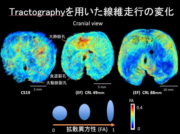
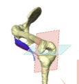
表紙に採用されました
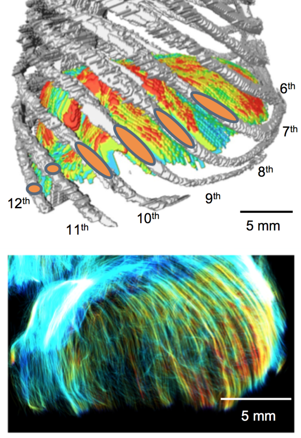
A precise understanding of human diaphragm development is essential in fetal medicine. To our knowledge, no previous study has attempted a three-dimensional (3-D) analysis and evaluation of diaphragmatic morphogenesis and development from the embryonic to the early fetal period. This study aimed to evaluate the morphogenesis and fibrous architecture of the developing human diaphragm during the late embryonic and early fetal periods. Fifty-seven human embryos and fetuses (crown-rump length [CRL] = 8–88 mm) preserved at the Congenital Anomaly Research Center of Kyoto University and Shimane University were analyzed. 3-D morphogenesis and fiber orientation of the diaphragm were assessed using phase-contrast X-ray computed tomography, T1-weighted magnetic resonance imaging (T1W MRI), and diffusion tensor imaging (DTI). T1W MR images and DTI scans were obtained using a 7-T MR system. The diaphragm was completely closed at Carnegie stage (CS) 20 and gradually developed a dome-like shape. The diaphragm was already in contact with the heart and liver ventrally in the earliest CS16 specimen observed, and the adrenal glands dorsally at CS19 or later. In the fetal period, the diaphragm contacted the gastric fundus in samples with a CRL ≥41 mm, and the spleen in samples with a CRL ≥70 mm. The relative position of the diaphragm with reference to the vertebrae changed rapidly from CS16 to CS19. The most cranial point of the diaphragm was located between the 4th and 8th thoracic vertebrae, regardless of fetal growth, in samples with a CRL ≥16 mm. Diaphragmatic thickness was nearly uniform (0.15–0.2 mm) across samples with a CRL of 8 mm to 41 mm. The sternal, costal, lumbar parts, and the area surrounding the esophageal hiatus thickened with growth in samples with a CRL ≥46 mm. The thickness at the center of the diaphragm and the left and right hemidiaphragmatic domes did not increase with growth. Tractography showed that the fiber orientation of the sternal, costal, and lumbar parts became more distinct as growth progressed in CS19 or later. All fibers in the costal and lumbar regions ran toward the left and right hemidiaphragmatic domes, except for those running to the caval opening and esophageal hiatus. The fiber orientation patterns from the right and left crura surrounding the esophageal hiatus were classified into three types. Distinct fiber directions between the costal and sternal, and between the costal and lumbar diaphragmatic parts were observable in samples with a CRL ≥46 mm. Anterior costal and sternal fibers ran toward the center. Fiber tracts around the center and the left and right hemidiaphragmatic domes; between the costal and lumbar orientations; and between the costal and sternal orientations showed a tendency for decreasing fractional anisotropy values with fetal growth, and showed less density than other areas. In conclusion, we used 3-D thickness assessment and DTI tractography to identify qualitative changes in the muscular and tendonous regions of the diaphragm during the embryonic and early fetal periods. This study provides information on normal human diaphragm development for the progression of fetal medicine and furthering the understanding of congenital anomalies.
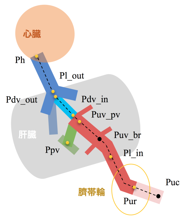
Three-dimensional analysis of the umbilical vein and the ductus venosus at the human embryonic and early fetal stages ヒト胚子期・胎児期初期における臍帯静脈と静脈管の3次元的解析 磯谷 菜穂子
67. Isotani N, Kanahashi T, Imai H, Yoneyama A, Yamada S, Takakuwa T. Regional differences in the umbilical vein and ductus venosus at different stages of normal human development. Anat Rec (Hoboken), 2024, 307, 3306-3326.DOI:10.1002/ar.25421
Morphogenesis of the pulmonary vein and the left atrial appendage in human embryos and early fetuses ヒト胚子・胎児期初期における肺静脈・左心耳の形態形成 福井 成美
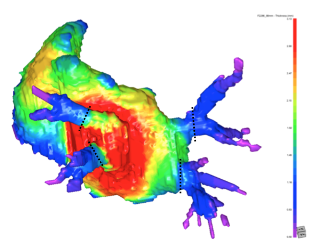
[Background] The left atrium (LA) forms the bulk of the cardiac fundus. Four pulmonary veins (PVs) flow from the fundus, and the left appendage (LAA) is joined to the anterior side via a stenosis. Toward the end of the fourth week of development, the common pulmonary vein (CPV) protrudes from the posterior wall of the LA. The CPV and the proximal portions of the four PVs dilate and are taken up by the LA wall, and finally, the four PVs protrude directly from the left atrium. The LAA originates from the primordial LA and forms during the fourth week of development. The adult LAA has one to four lobe-like structures, and the interior of the LAA has a comb-like structure created by pectinate muscles. The time of completion of the PV uptake into the LA wall has been controversial in previous studies, and morphogenesis of the LAA has been reported only in adult cases. We provided the data showing the PV uptake process and the LAA development based on high-resolution image data and three-dimensional information. [Materials & Methods] Twenty-four human embryos and twenty fetuses were selected for this study. The PV was observed on CT images obtained by phase CT imaging and MR images obtained by high-resolution MRI imaging. 3D images of the heart portion were reconstructed based on the MR images, and morphological observation and quantitative evaluation of the PVs and LAA were performed. [Results] The number of PV was one from CS 17 to CS 18, two or four from CS 18 to CS 21, and three or four after CS 22. In specimens with two or more PVs, the distance between the left and right PVs increased with CRL, as did the reconstruction of the LA thickness between the left and right-sided PV. The distance between the superior and inferior PVs was closer than between the left and right-sided PVs. When there were two PVs from CS18 to CS 21, they flowed into the LA from the dorsal side; when there were three or four PVs, they entered tangentially into the dorsal part of the LA from the lateral to medial direction. Regarding the cross-sectional area of the PV, the LSPV was the smallest, while the other three PVs were similar. In shape, the LSPV was the most flattened; the other three PVs were similar. The LAA thickness was thickest near the center, and it became radially thinner from there. The LAA orifice increased in area and tended to become more flattened with CRL. [Conclusion] The PV uptake is thought to begin around CS 18 and complete from CS 18 to CS 22. The proximal portion of the LSPV is smaller in cross-sectional area and circumference than the other three PVs and is more flattened. Four PVs enter tangentially into the dorsal part of the LA from the lateral to medial direction. And the distance between the superior and inferior PV is closer than between the left and right-sided PV. The LAA thickness is thickest near the center, and it becomes radially thinner from there. The LAA orifice is found to increase in area and become more flattened with CRL.
61. Fukui N, Kanahashi T, Matsubayashi J, Imai H, Yoneyama A, Otani H, Yamada S, Takakuwa T. Morphogenesis of the pulmonary vein and left atrial appendage in human embryos and early fetuses. J Anatomy 2023, in press, https://doi.org/10.1111/joa.13941
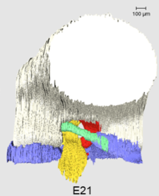
Three-dimensional imaging analysis of developmental process of posterior meniscofemoral ligament in rat embryo ラット胎仔における後半月大腿靭帯の発生機序の三次元的解析 石田かのん
Objectives: The posterior meniscofemoral ligament (pMFL) of the knee joint has been reported to contribute to knee joint stability and to be associated with the discoid lateral meniscus (DLM); however, its developmental process in healthy knees has not been studied. In this study, we analyzed the developmental process using three-dimensional (3D) reconstructed images in addition to two-dimensional observations. Owing to ethical constraints, rats were selected for this study because of the advantage of their similar knee structure to humans and the availability of multiple fetuses. The purpose of this study was to analyze pMFL development in rat knee joints three-dimensionally and examine its relationship with other knee joint components. Methods: The shape and position of hindlimbs of Wistar rats at E16-E21 were confirmed with HE-stained tissue sections. Serial episcopic fluorescence images of the hindlimbs of E17-E21 were respectively captured by episcopic fluorescence image capture (EFIC), from which 3D images were reconstructed using Amira software. The pMFL length, deflection, angle, and volume were measured in 3D images. The volumes of the anterior cruciate ligament (ACL), posterior cruciate ligament (PCL), and menisci were also measured and the ratio of the volume to the mean value at E17 of each component, including pMFL, was compared.Results: pMFL was observed from E17 and was attached to the medial femoral condyle and lateral meniscus at all stages. The pMFL length and volume of each knee joint component increased significantly between E19 and E21; no significant variation was observed in the pMFL deflection and angle throughout all phases. The Volume ratios showed that all components showed similar increasing trends until E19, but the menisci and PCL increased significantly from E20. Discussion: While the length of pMFL and volume of each component increased significantly after E19, there was little variation in angle throughout the stages studied, suggesting that the pMFL and surrounding components developed with a positional relationship. A higher attachment position of the pMFL to the femur may cause DLM , and the present results may help in understanding the mechanism of DLM development. When compared with a report that hindlimb movement in rats increases between E16 and E19, the time when the length of pMFL and volume of each component significantly increased is just after this, indicating that the developmental process is divided into two phases. As rat knees are loaded differently from human knees, further studies are required on the developmental process of human knees. Conclusion: The developmental process of pMFL and knee joint components in rat embryos was analyzed three-dimensionally. This study improves our understanding of the developmental processes of the normal pMFL and knee joint components.
Tanima M, Ishida K, Ishikawa A, Yamada S, Takakuwa T, Aoyama T, Three-dimensional imaging analysis of the developmental process of posterior meniscofemoral ligaments in rat embryos. Cells Tissues Organs 2024, in press, DOI: 10.1159/000536108
Nohara A, Owaki N, Matsubayashi J, Katsube M, Imai H, Yoneyama A, Yamada S, Kanahashi T, Takakuwa T. Morphometric analysis of secondary palate development in human embryos. J Anatomy, 2022, 241(6), 1287-1302, 2022, DOI:10.1111/joa.13745
Abstract
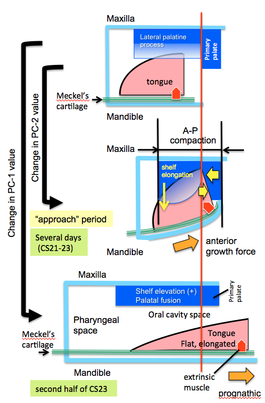
Rapid shelf elevation and contact of the secondary palate and fusion reportedly occur due to a growth-related equilibrium change in the structures within the oro-nasal cavity. This study aimed to quantitatively evaluate complex three-dimensional morphological changes and their effects on rapid movements, such as shelf elevation and contact, and fusion. Morphological changes during secondary palate formation were analyzed using high-resolution digitalized imaging data (phase-contrast X-ray computed tomography and magnetic resonance images) obtained from 22 human embryonic and fetal samples. The three-dimensional images of the oro-nasal structures, including the maxilla, palate, pterygoid hamulus, tongue, Meckel’s cartilage, nasal cavity, pharyngeal cavity, and nasal septum, were reconstructed manually.
palatal shelves were not elevated in all the samples at Carnegie stage (CS)21 and CS22 and in three samples at CS23. In contrast, the palatal shelves were elevated but not in contact in one sample at CS23. Further, the palatal shelves were elevated and fused in the remaining four samples at CS23 and all three samples from the early fetal period. For each sample, 70 landmarks were subjected to Procrustes and principal component (PC) analysis. PC-1 accounted for 67.4% of the extracted gross changes before and after shelf elevations. Notably, the PC-1 values of the negative and positive value groups differed significantly. The PC-2 value changed during the phases in which the change in the PC-1 value was unnaturally slow and stopped at CS22 and the first half of CS23. This period, defined as the “approach period”, corresponds to the time before dynamic changes occur as the palatal shelves elevate, the tongue and mandibular tip change their position and shape, and secondary palatal shelves contact and fuse. During the “approach period”, measurements of PC-2 changes showed that structures on the mandible (Meckel’s cartilage and tongue) and maxilla (palate and nasal cavity) did not change positions, albeit both groups of structures appeared to be compressed anterior-posteriorly. However, during and after shelf elevation, measurements of PC-1 changes showed significant changes between maxillary and mandibular structures, particularly positioning of the shelves above the tongue and protrusion of the tongue and mandible. These results suggest an active role for Meckel’s cartilage growth in repositioning the tongue to facilitate shelf elevation. The present data representing three distinct phases of secondary palate closure in humans can advance the understanding of morphological growth changes occurring before and after the horizontal positioning of palatal shelves and their fusion to close the secondary palate in humans successfully.
石川さんが、第9回 International Symposium on Regenerative Rehabilitationで発表しました(2022年10月27-29日)
Ishikawa A, Nagai-Tanima M, Ishida K, Imai H, Yamada S, Aoyama T, Takakuwa T. Three-dimensional morphological comparison of the knee at different stages of normal human development. the 9th Annual International Symposium on Regenerative Rehabilitation 2022年10月27-29日、於:Austin,TEXAS.