


発達神経科学会第3回学術集会で発表しました。(2014.10.16-17; 東京)
ヒト胚子における Willis 動脈輪の形成について
小池哲平、山田重人、上部千賀子、高桑徹也
ヒト胚子期における脳形態形成の解析
白石直樹、片山愛里、中島 崇、上部千賀子、巨瀬 勝美、山田 重人、高桑 徹也
|
||||
  |
発達神経科学会第3回学術集会で発表しました。(2014.10.16-17; 東京) ヒト胚子における Willis 動脈輪の形成について 小池哲平、山田重人、上部千賀子、高桑徹也 ヒト胚子期における脳形態形成の解析 白石直樹、片山愛里、中島 崇、上部千賀子、巨瀬 勝美、山田 重人、高桑 徹也 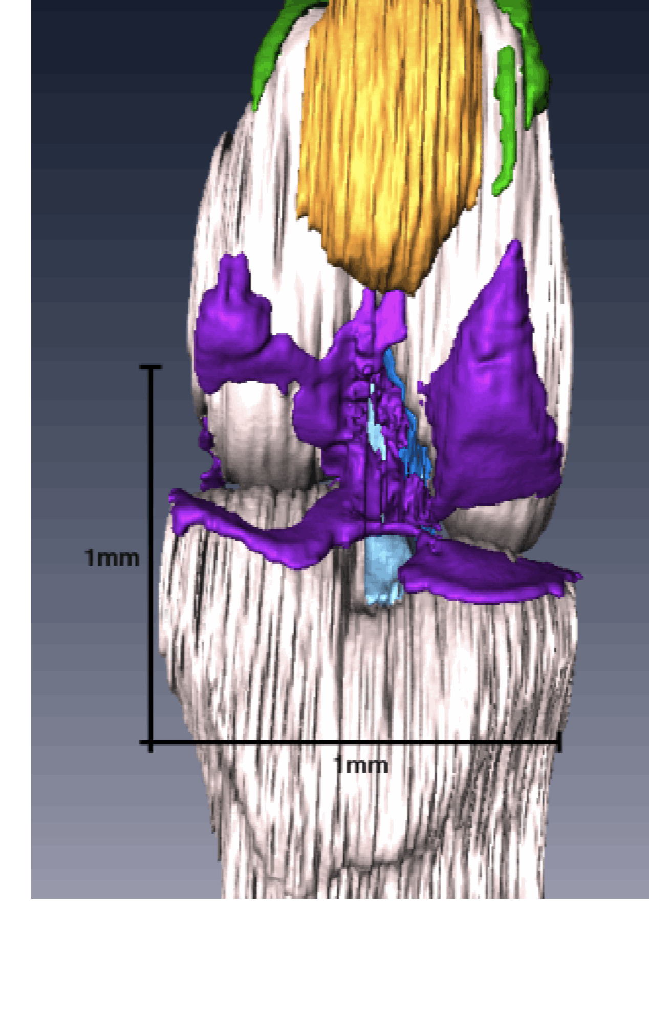 高石くんの卒業論文「膝関節の形態形成; EFICを用いた3次元的解析」がOsteoarthritis Cartilage ( 特集号Imaging in Osteoarthritis)に掲載されました。 9. Takaishi R, Aoyama T, Takakuwa T, et al; Three-dimensional reconstruction of rat knee joint using episcopic fluorescence image capture, Osteoarthritis Cartilage, 2014; 22(10), 1401-1409. 10.1016/j.joca.2014.06.016
SummaryObjectiveDevelopment of the knee joint was morphologically investigated, and the process of cavitation was analyzed by using episcopic fluorescence image capture (EFIC) to create spatial and temporal three-dimensional (3D) reconstructions. MethodsKnee joints of Wister rat embryos between embryonic day (E)14 and E20 were investigated. Samples were sectioned and visualized using an EFIC. Then, two-dimensional image stacks were reconstructed using OsiriX software, and 3D reconstructions were generated using Amira software. ResultsCavitations of the knee joint were constructed from five divided portions. Cavity formation initiated at multiple sites at E17; among them, the femoropatellar cavity (FPC) was the first. Cavitations of the medial side preceded those of the lateral side. Each cavity connected at E20 when cavitations around the anterior cruciate ligament (ACL) and posterior cruciate ligament (PCL) were completed. ConclusionCavity formation initiated from six portions. In each portion, development proceeded asymmetrically. These results concerning anatomical development of the knee joint using EFIC contribute to a better understanding of the structural feature of the knee joint. |
|||