豊田さんの卒業研究がAnatomical Recordに掲載されました。おめでとうございます。
- CS17から胎児期初期の内耳(膜迷路)の形態形成を検討
- 耳小胞は背腹軸に沿って伸び、
- CS 17 ;終末リンパ付属器と蝸牛管 (CD) に分化
- CS18; CDのらせんの開始、前後SDが共通脚を形成
- post-embryonic phase; CDは 2 回以上のターン立体的
- 前部、後部、および横方向の SD の長さの直線的な増加がこの順序で観察され、CD の長さは発達の過程で指数関数的に増加。
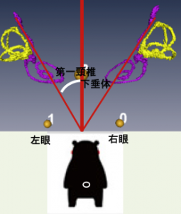
- 内耳の位置は、観察期間中一定
- 外側半規管の体軸に対する角度は成人と異なる、成長に伴い内耳の向きが変化することを示唆
17. Toyoda S, Shiraki N, Yamada S, Uwabe C, Imai H, Matsuda T, Yoneyama A, Takeda T, Takakuwa T, Morphogenesis of the inner ear at different stages of normal human development. Anatomical Record, 298:2081–2090 (2015), doi: 10.1002/ar.23268
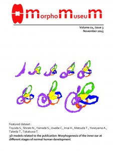
作成した立体データの代表的なものはMorphoMuseumに発表されました。
Toyoda S, Shiraki N, Yamada S, Uwabe C, Imai H, Matsuda T, Yoneyama A, Takeda T, Takakuwa T, Morphogenesis of the human inner ear membranous labyrinth. MorphoMuseuM 1 (3)-e6. doi: 10.18563/m3.1.3.e6
ABSTRACT
This study examined the external morphology and morphometry of the human embryonic inner ear membranous labyrinth and documented its three-dimensional position in the developing embryo using phase-contrast X-ray computed tomography and magnetic resonance imaging. A total of 27 samples between Carnegie stage (CS) 17 and the postembryonic phase during trimester 1 (approximately 6–10 weeks after fertilization) were included. The otic vesicle elongated along the dorso-ventral axis and differentiated into the end lymphatic appendage and cochlear duct (CD) at CS 17. The spiral course of the CD began at CS18, with anterior and posterior semicircular ducts (SDs) forming prominent circles with a common crus. The spiral course of the CD comprised more than two turns at the postembryonic phase, at which time the height of the CD was evident. A linear increase was observed in the length of anterior, posterior, and lateral SDs, in that order, and the length of the CD increased exponentially over the course of development. Bending in the medial direction was observed between the cochlear and vestibular parts from the latero-caudal view, with the angle decreasing during development. The position of the inner ear was stable throughout the period of observation on the lateral to ventral side of the rhombencephalon, caudal to the pontine flexure, and adjacent to the auditory ganglia. The plane of the lateral semicircular canal was approximately 8.0°–14.6° with respect to the cranial caudal (z-)axis, indicating that the orientation of the inner ear changes during growth to adulthood.