第59回先天異常学会で、発表しました(2019.7.26-28, 名古屋)The 13th World Congress of the International Cleft Lip and Palate Foundation -CLEFT 2019-と合同開催でした。
Nohara A, Owaki N, Manesco C, Katsube M, Yamada S, Imai H, Matsuda T, Yoneyama A, Takakuwa T, Relationship between fusion of lateral palatal shelves and growth of Mandible (Meckel’s cartilage)
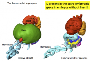
PUH occurred independently of liver
Takakuwa T. Intestinal loop formation: herniation into the extraembryonic coelom and return to the abdominal coelom (招待講演)
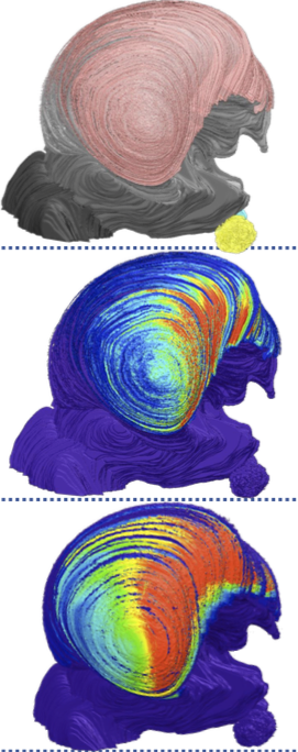
Drastic changes occur during the development of the intestinal loop (IL), including physiological umbilical herniation (PUH) and its return. The present study was designed to analyze such developments three-dimensionally during human embryonic and early fetal period.
Materials and Methods: The software AMIRA was used to analyze the 3D digitalized data (high-resolution MRI, phase-contrast X-ray CT) obtained from the Kyoto Collection.
Results and Discussion: Based on the results of our analysis, the following time line and main features of IL formation were revealed:
Herniate phase (Carnegie stage (CS)14-CS23, Crown-rump length (CRL) < 35 mm): IL rotation was initially observed as a slight deviation of the duodenum and colorectum from the median plane up to CS16. The PUH was noticeable after CS16. The IL displayed a hairpin-like structure, with the superior mesenteric artery (SMA) running parallel to the straight part and the cecum located to the left at CS18. The IL rotated around the SMA only during the early stages (until CS19). The IL gradually moved away, running transversely after CS19. Embryos with liver malformation showed PUH, which indicated that PUH occurred independent of liver volume.
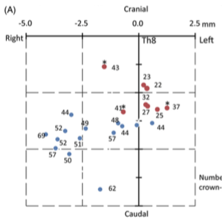
Transition phase (CRL = 37, 41, and 43 mm): Intestinal return began from proximal to distal part in samples with CRL of 37 mm. The cecum returned before the distal end of the small intestine (ileum) in samples with CRLs of 41 and 43 mm.
Return phase: The cecum immediately reached its final position in the right lower quadrant of the abdomen (the adult position). The anti-clockwise “en-bloc rotation” described by descent and fixation of the cecum in the abdominal cavity may not exist. A rapid increase in the space available for the intestine in the abdominal coelom that exceeded the intestinal volume in the extraembryonic coelom was observed. The height of the umbilical ring increased in a stepwise manner between the transition and return phases and its height in the return phase was comparable to or higher than that of the hernia tip during the herniation phase. We speculated that the space is generated to accommodate the herniated portion of the intestine, similar to the intestine wrapping into the abdominal coelom as the height of the umbilical ring increases.
Conclusion: The data obtained in the present study demonstrate the precise timeline of IL formations, which indicate several points of discrepancy in the results of previous studies.
42. Nagata A, Hatta S, Imai H, Yamada S, Takakuwa T. Position of the cecum in the extraembryonic and abdominal coelom in the early fetal period. Congenit Anom 2020, 60 (3) 87-88. doi: 10.1111/cga.12348.