アクセスカウンター

2019年10月
 123456
78910111213
14151617181920
21222324252627
28293031  

日本人類学会で発表

第73回日本人類学会で発表しました(2019.10.12-14、佐賀)。

われわれの研究も人類の研究なので、発表してみました。どの程度受け入れられたでしょうか。

ヒト胚子期における気管支分岐形成の三次元的定量解析
藤井 瀬菜、村中 太河、松林 潤、米山 明男、兵藤 一行、山田 重人、高桑 徹也
ヒト胚子期・胎児期初期における気管・軟骨の3次元的解析
山崎 優、藤井 瀬菜、石川 葵、山田 重人、高桑 徹也
Carnegie Stage (CS) でのヒト後腎における腎小体の発生段階と立体的分布
北沢 遥、石山 華、石川 葵、松林 潤、山田 重人、高桑 徹也

腎臓の回転、移動についての論文がAnat Recに掲載

腎臓の回転、移動についての論文がAnat Recに受諾されました。

腎臓が発生時に上昇し回転することは教科書に書かれています。その動きを3Dでしっかり観察したこと、集合管尿路系の形成との関連で論じたことが特徴です。

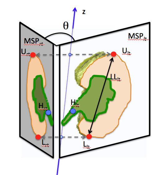
  • 腎門の向きは、CS17 (34.4  度) と 18 (122.3度) の間で大幅に変化
  • 尿路樹の分岐数が CS17 での1.61 から CS18で3.20 に増加
  • 枝の数の増加は、後腎の成長と腎門の方向に影響を与えた。

石山さんの修士の仕事に石川さんがデータを追加してくれました。

37. Ishiyama H, Ishikawa A, Imai H, Matsuda T, Yoneyama A, Yamada S, Takakuwa T. Spatial relationship between the metanephros and adjacent organs according to the Carnegie stage of development. Anat Rec 2019. 302, 1887-2104. DOI: 10.1002/ar.24103

ABSTRACT

The morphological changes in the metanephros and its spatial relationship to the adjacent organs was evaluated based on the Carnegie stages (CSs) from 14 through 23. The imaging modalities used included magnetic resonance imaging (N = 4), phase-contrast X-ray computed tomography (N = 11), and serial histological sections (N = 40), supplemented by three-dimensional image reconstruction. The orientation of the hilus of the metanephros changed significantly between CS17 (34.4 ± 13.7 degrees) and 18 (122.3 ± 38.1 degrees), with an increase in the number of branches of the urinary collecting system, from 1.61 ± 0.42 at CS17 to 3.20 ± 0.35 at CS18. This increase in the number of branches influenced the growth of the metanephros and the orientation of its hilus. The right and left metanephroses were in proximity throughout the embryonic period. The local maximum interpole distances were observed at CS18 (0.87 ± 0.11 mm for the upper and 0.50 ± 0.25 mm for the lower pole). Mesenchymal tissue was observed between the metanephros and iliac arteries, as well as between the right and left metanephros. Throughout development, the position of the lower pole of the metanephros remained adjacent to the aortic bifurcation. The position of the upper pole, referenced with respect to the aortic bifurcation, increased by >2.0 mm, reflecting the longitudinal growth of the metanephros. Our findings provide a detailed description of the morphogenesis of the metanephros and of its hilus, which might contribute to our understanding of congenital malformations and malpositions of the kidneys. 

肝臓形成不全でも生理的臍帯ヘルニアは起きる: Anat Recに掲載

胚子期、消化管は大きい肝臓に押し出され臍帯内に脱出すると言われています。この機序は、発生の大家Mall博士が19世紀末に提唱し、今多くの教科書に記されています。そうしたら、肝臓形成不全で肝臓が小さい胚子、肝臓がない胚子ではどうなのでしょうか? 

  • 本研究では、消化管臍帯ヘルニアが肝臓の容積に関係なく発生することを示す標本を複数提示することで、発生の大家の提示した説に異議を唱えました。

Kanahashi T, Yamada S, Yoneyama A, Takakuwa T. Relationship Between Physiological Umbilical Herniation and Liver Morphogenesis During the Human Embryonic Period: A Morphological and Morphometric Study. Anat Rec 2019, 302, 1968-1976. doi: 10.1002/ar.24149.

肝臓無形成でも生理的臍帯ヘルニアがみられる

ABSTRACT

It is widely hypothesized that physiological umbilical herniation (PUH) in humans occurs, because the liver occupies a large space in the abdominal cavity, which pushes the intestine into the extraembryonic coelom during the embryonic period. We have recently shown the presence of the intestinal loop in the extraembryonic coelom in embryos with liver malformation. Here, we analyzed the relationship between the liver and the PUH at Carnegie stage 21 of four embryos with liver malformation, including two with hypogenesis (HY1, HY2) and two with agenesis (AG1, AG2), using phase-contrast X-ray computed tomography and compared them with two control embryos. The intestinal loop morphology in the malformed embryos differed from that in the control embryos, except in HY1. The length of the digestive tract in the extraembryonic coelom of the embryos with liver malformation was similar to or longer than that of the controls. The rate of intestinal loop lengthening in the extraembryonic coelom compared with that of the total digestive tract in all embryos with liver malformation was similar to or higher than that of the controls. The estimated total abdominal cavity volume in the embryos with liver malformation was considerably smaller than that of the controls, while the intestinal volume was similar. The cardia and proximal portion of the pancreas connecting to the duodenum were located at almost identical positions in all the embryos, whereas other parts of the upper digestive tract deviated in the embryos with abnormal livers. Thus, our results provided evidence that PUH occurred independently of liver volume.