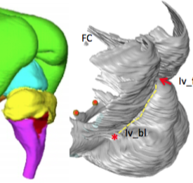
松成さんの卒業研究がAnat Recに掲載されました。
硬膜のような袋状の構造物は、組織形態学的手法では立体的な形状がとらえにくかったのですが、MRI等の立体情報の活用により可能になりました。
- 胚子期から胎児期の64個体の小脳テントの形成について、脳の形態との相互関係から立体的に解析
- 小脳テントの特徴により、胚子期、胎児期初期、中期にわけられる
- 大脳の後方への成長、中脳屈曲の解消等の脳形成時の変化がテントの形状や位置に影響をあたえる
59. Matsunari C, Kanahashi T, Otani H, Imai H, Yamada S, Okada T, Takakuwa T. Tentorium cerebelli formation during human embryonic and early fetal development. Anat Rec (Hoboken) 2023, 306(3), 515-526
Abstract
The morphologies of the fetal tentorium cerebelli (TC) and brain influence each other during development. This study aimed to analyze and more comprehensively understand the three-dimensional morphogenesis of the TC and fetal brain. We examined magnetic resonance imaging from 64 embryonic and fetal specimens (crown-rump length range, 9.2–225 mm). During the embryonic period, the lateral folds of the TC elongated to traverse the middle part of the midbrain. The TC and falx cerebri appeared separated, and no invaginations at the parieto-occipital region were observed. In the early fetal period, the cerebrum covered approximately half of the midbrain. The separation of the dural limiting layer at the parieto-occipital region widened from the posterior cerebrum to the cranial cerebellum. The lateral folds of the TC were spread between its tip, continuous with the falx cerebri, and its base plane, located between the midbrain and rostral hindbrain. Differences in the TC components’ growth directions gradually diminished as the cerebrum covered the midbrain. We observed rotation of the TC at its median section according to its growth, which ceased in the middle fetal period. The brainstem and cerebellum extended inferiorly via differential growth, with the cerebrum covering them superiorly. The morphology of the TC curved to conform to the cerebellar and cerebral surfaces. Our present study suggests that factors affecting TC morphology differ between the early and middle fetal periods. Present data provided a more comprehensive view of TC formation according to developmental stage.


