


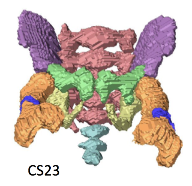
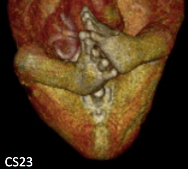
胚子期の股関節の肢位についての論文がPLoS Oneに掲載されました。熊野くんが卒業研究で行ったデータに、胚子期の標本のデータを追加し解析しました。大腿骨(股関節)の肢位を正確に測定し、下肢の発達の時間軸を確立することを目的とした論文です。
- CS19-23の胚子期157体と胎児(CRL:37.2-225mm)18体のMRI画像を対象とした。
- 下肢と骨盤の選択された8つのランドマークの3次元座標を用いて大腿骨の肢位を計測。
- 胚子期には、股関節の屈曲、外転、側転の3つの姿勢パラメータが互いに線形相関を示し、各段階の大腿骨姿勢は3次元的に一定で、成長に応じて緩やかで滑らかな変化を示すことが示唆された。
60. Takakuwa T, Saizonou MA, Fujii S, Kumano Y, Ishikawa A, Aoyama T, Imai H, Yamada S, Kanahashi T. Femoral posture during embryonic and early fetal development: An analysis using landmarks on the cartilaginous skeletons of ex vivo human specimens. PLOS one, 2023, 18(5): e0285190. https://doi.org/10.1371/journal.pone.0285190.
Abstract
The pre-axial border medially moves between the fetal and early postnatal periods, and the foot sole can be placed on the ground. Nonetheless, the precise timeline when this posture is achieved remains poorly understood. The hip joint is the most freely movable joint in the lower limbs and largely determines the lower-limb posture. The present study aimed to establish a timeline of lower-limb development using a precise measurement of femoral posture. Magnetic resonance images of 157 human embryonic samples (Carnegie stages [CS] 19–23) and 18 fetal samples (crown rump length: 37.2–225 mm) from the Kyoto Collection were obtained. Three-dimensional coordinates of eight selected landmarks in the lower limbs and pelvis were used to calculate the femoral posture. Hip flexion was approximately 14° at CS19 and gradually increased to approximately 65° at CS23; the flexion angle ranged from 90° to 120° during the fetal period. Hip joint abduction was approximately 78° at CS19 and gradually decreased to approximately 27° at CS23; the average angle was approximately 13° during the fetal period. Lateral rotation was greater than 90° at CS19 and CS21 and decreased to approximately 65° at CS23; the average angle was approximately 43° during the fetal period. During the embryonic period, three posture parameters (namely, flexion, abduction, and lateral rotation of the hip) were linearly correlated with each other, suggesting that the femoral posture at each stage was three-dimensionally constant and exhibited gradual and smooth change according to growth. During the fetal period, these parameters varied among individuals, with no obvious trend. Our study has merits in that lengths and angles were measured on anatomical landmarks of the skeletal system. Our obtained data may contribute to understanding development from anatomical aspects and provide valuable insights for clinical application.