


福井さんの修士論文がJ Anatomyに受諾されました。ヒト胎児期初期の左房の形成を肺静脈のとりこみと左心耳の形成を中心に解析しました。
- 23例の胚子期、19例の胎児期初期標本を選択した。
- 高分解能イメージング(位相CTとMRI)から三次元心臓画像を再構成し、左心耳を含む肺静脈と左心房を形態学的および定量的に評価した。
- 心臓の背側中膜結合が退縮したとき(CS18以降)、心膜折り返しの位置が2つの肺静脈(右肺静脈と左肺静脈)と4つの肺静脈でほぼ同時に決定された。
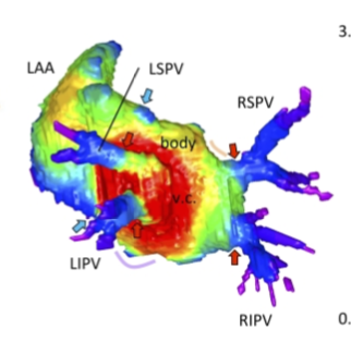
- 左心房のひだが体部と静脈成分の接合部に存在することが確認された。
- 左心房の肺静脈由来の静脈成分は成長に比例して増加することが示された。肺静脈成分と左心房体との接合部は徐々に目立たなくなったが、観察された初期胎児期の終わりにはまだ認識可能であった。
Fukui N, Toru KanahashiT, MatsubayashiJ, ImaiH, YoneyamaA, OtaniH, YamadaS, Takakuwa T. Morphogenesis of the pulmonary vein and left atrial appendage in human embryos and early fetuses. J Anatomy 2024, 244, 142-158, in press, https://doi.org/10.1111/joa.13941
Abstract
The left atrium wall has several origins, including the body, appendage, septum, atrial–ventricular canal, posterior wall, and venous component. Here, we describe the morphogenesis of left atrium based on high-resolution imaging (phase-contrast X-ray computed tomography and magnetic resonance imaging). Twenty-three human embryos and 19 fetuses were selected for this study. Three-dimensional cardiac images were reconstructed, and the pulmonary veins and left atrium, including the left atrial appendage, were evaluated morphologically and quantitatively. The positions of the pericardial reflections were used as landmarks for the border of the pericardial cavity. The common pulmonary vein was observed in three specimens at Carnegie stage 17–18. The pericardium was detected at the four pulmonary veins (left superior, left inferior, right superior, and right inferior pulmonary veins) at one specimen at Carnegie stage 18 and all larger specimens, except the four samples. Our results suggest that the position of the pericardial reflections was determined at two pulmonary veins (right and left pulmonary vein) and four pulmonary veins almost simultaneously when the dorsal mesocardial connection between the embryo and heart regressed. The magnetic resonance images and reconstructed heart cavity images confirmed that the left atrium folds were present at the junction between the body and venous component. Three-dimensional reconstruction showed that the four pulmonary veins entered the dorsal left atrium tangentially from the lateral to the medial direction. More specifically, the right pulmonary veins entered at a greater angle than the left pulmonary veins. The distance between the superior and inferior pulmonary veins was shorter than that between the left and right pulmonary veins. Three-dimensional reconstruction showed that the venous component increased proportionally with growth. No noticeable differences to discriminate between the right and left parts of the venous component emerged, while the junction between the venous component and body gradually became inconspicuous but was still recognizable by the end of the observed early fetal period. The left superior pulmonary vein had the smallest cross-sectional area and most flattened shape, whereas the other three were similar in area and shape. The left atrial appendage had a large volume in the center and extended to the periphery as a lobe-like structure. The left atrial appendage orifice increased in the area and tended to become flatter with growth. The whole left atrium volume^(1/3) increased almost proportionally with growth, parallel to the whole heart volume. This study provided a three-dimensional and quantitative description of the developmental process of left atrium, comprising the venous component and left atrial appendage formation, from the late embryonic to the early fetal stages.