


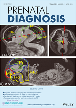
小林さんの卒論がPrenatal Diagnosisに掲載されました。ヒト胚子期の標本を用いて脳の発生に伴う計測値の変化、特徴を論じたものです。
- 胚子期の標本の脳の7直線、3領域の面積、体積を検討
- すべての直線計測値はCRLと相関
- bitemporal lengthと全脳、大脳体積は高い相関
得られた結果は、超音波データの修正と比較、出生前診断の改善に貢献することが期待されます。
また、同号の表紙にも採用されました。
23. Kobayashi A, Ishizu K, Yamada S, Uwabe C, Kose K, Takakuwa T, Morphometric human embryonic brain features according to developmental stage, Prenatal Diagnosis, 36:338–345, 2016, DOI: 10.1002/pd.4786. DOI: 10.1002/pd.4818
Abstract
Objectives
The present study investigated linear, area, and volume measurements of human brain samples according to Carnegie stages (CS) in an attempt to select suitable morphometric features that reflect embryonic development.
Methods
Using magnetic resonance imaging, we measured seven linear segments, three separate areas, and three regional volumes in 101 samples between CS13 and 23. Brain volume was determined via manual segmentation of the magnetic resonance image, whereby a formula was generated to estimate the volume of each linear measurement.
Results
All parameters correlated with crown-rump length. Bitemporal length and mesencephalic height increased linearly according to the CS, and a high correlation between bitemporal length and both whole-brain (r = 0.98) and prosencephalon (r = 0.99) volumes was found when brain cavity volume was excluded.
Conclusion
Morphometric data related to human embryonic stages are valuable for correcting and comparing sonographic data. The present approach may contribute to improvements in prenatal diagnostics by enabling the selection of more suitable measurements during early embryonic stages.