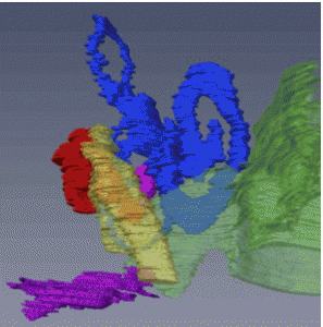
尾関さんの修士論文のうち、中耳耳小骨の形成と外耳、内耳との立体的位置関係についてAnat Recに掲載されました。
MEO の形成と外耳と内耳の接続のタイムラインを決定しました。
- 軟骨頭蓋は、CS18 までに認識可能
- 軟骨形成開始は耳小骨(MEO)間で若干の相違。
- CS19;槌骨、キヌタ骨、アブミ骨の順序で、前後方向に配置 CS22以降;MEO同士が関節を介し接続。
- CS21以降にすべてのMEOの軟骨形成を確認
- CS23;アブミ骨基底部分はfoot plate状ではない
- ツチ骨のハンドルは 外耳道からは離れている
- 耳介軟骨と耳嚢の軟骨膜境界は明確
26. Ozeki-Satoh M, Ishikawa A, Yamada S, Uwabe C, Takakuwa T. Morphogenesis of the Middle Ear Ossicles and Spatial Relationships with the External and Inner Ears during the Embryonic Period, Anat Rec 299:1325–1337, 2016, DOI 10.1002/ar.23457
Abstract
We describe the three-dimensional morphogenesis of the middle ear ossicles (MEOs) according to Carnegie stage (CS) in human embryos. Seventeen samples including 33 MEOs from CS18 to 23 were selected from the Kyoto Collection. The primordia of the MEOs and related structures were histologically observed and three-dimensionally reconstructed from digital images. The timing of chondrogenesis was variable among structures. The stapes was recognizable as a vague condensation of the mesenchymal cells in all samples from CS18, whereas the malleus and incus were recognizable at CS19. Chondrogenesis of all MEOs was evident in all samples after CS21. The chondrocranium was recognizable in all samples by CS18, and the perichondrium border of the auricular cartilage and otic capsule was distinct in all samples at CS23. At CS19, the MEOs were positioned in the anterior to posterior direction, following the order malleus, incus, stapes, which adjusted gradually during development. The MEOs connected in all samples after CS22. The stapes was located close to the vestibular part of the inner ear, although the basal part was not differentiated into the “footplate” form, even at CS23. The handles of the malleus were close to the tubotympanic recess at CS23, but were distant from the external auditory meatus. Determining the timeline of the formation of MEOs and connection of the external and inner ears can be informative for understanding hearing loss caused by failure of this connection. These data may provide a useful standard for morphogenesis, and will contribute to distinguishing between normal and abnormal MEO development.


