


石川さんの論文がAnatmial Recordに掲載されました。
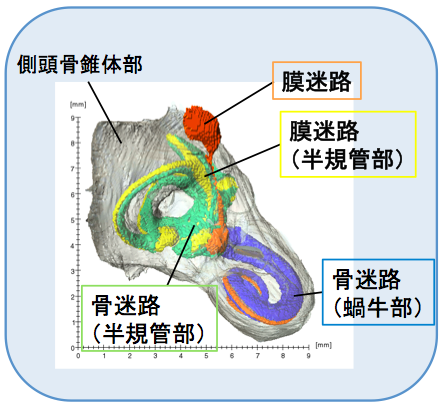
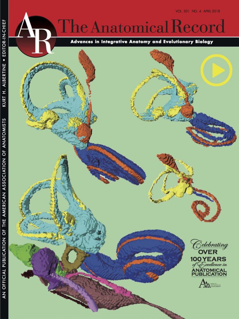
内耳のうち膜迷路、骨迷路の間にあるperiotic spaceの形成に着眼したユニークな論文です。図が表紙に採用されました!!
- 膜迷路は、CS23のサンプルで判別可能
- CRL 14.4–197 mmの観察期間中に長さは8 倍以上に直線的に成長
- periotic spaceは、CRL35 mm の標本で蝸牛の前庭と基底部周囲で最初に検出
- 115 mm CRL で膜迷路をほぼ覆う。
- タイムテーブルに従って、膜迷路、periotic space、otic capsuleの骨化が連続して発生
32. Ishikawa A, Ohtsuki S, Yamada S, Uwabe C, Imai H, Matsuda T, Takakuwa T. Formation of the periotic space during the early fetal period in humans, Anat Rec, 2018, 301(4);563-570, 10.1002/ar.23764, 10.1002/ar.23657
Abstract
The inner ear is a very complicated structure, composed of a bony labyrinth (otic capsule; OC), membranous labyrinth, with a space between them, named the periotic labyrinth or periotic space. We investigated how periotic tissue fluid spaces covered the membranous labyrinth three-dimensionally, leading to formation of the periotic labyrinth encapsulated in the OC during human fetal development. Digital data sets from magnetic resonance images and phase-contrast X-ray tomography images of 24 inner ear organs from 24 human fetuses from the Kyoto Collection (fetuses in trimesters 1 and 2; crown—rump length: 14.4–197 mm) were analyzed. The membranous labyrinth was morphologically differentiated in samples at the end of the embryonic period (Carnegie stage 23), and had grown linearly to more than eight times in size during the observation period. The periotic space was first detected at the 35-mm samples, around the vestibule and basal turn of the cochlea, which elongated rapidly to the tip of the cochlea and semicircular ducts, successively, and almost covered the membranous labyrinth at the 115-mm CRL stage or later. In those samples, several ossification centers were detected around the space. This article thus demonstrated that formation of the membranous labyrinth, periotic space (labyrinth), and ossification of the OC occurs successively, according to an intricate timetable.