


ヒト胎児期初期の脳の形態形成についての論文がJ Anatomyに掲載されました。5年以上かけて、ようやくまとめることができました。
- 高解像度MRIを用いて胎児期初期の(受胎後 9 ~ 13 週) の脳の形態計測学的分析を行った。
- 脳梁の出現(CRL62mm), 皮質内側の脳溝の出現(CRL114mm),
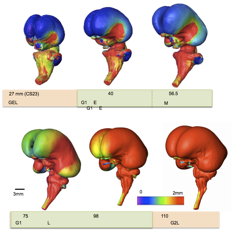
- 小脳半球、後外側裂溝、小脳半分の結合、虫部の形成(CRL43.5mm), 一次裂溝の出(CRL56mm), 錐体前裂(CRL75mm),
- 大脳側面における大脳の比率 (長さ/高さ) と基準線に対する大脳の角度は、大脳の成長と C 字型の形成を反映している可能性がある。
- これらの所見は、胎児期初期を細分化するための候補となる可能性がある。
52. Takakuwa T, Shiraishi N, Terashima M,Yamanaka M, Okamoto I, Imai H, Ishizu K, Yamada S, Ishikawa A, Kanahashi T. Morphology and morphometry of the human early fetal brain: A three-dimensional analysis. J Anatomy; 239 (2) 498-516, 2021, doi.org/10.1111/joa.13433
Abstract
Morphometric analyses in the early foetal phase (9-13 postconceptional week) are critical for evaluating normal brain growth. In this study, we assessed sequential morphological and morphometric changes in the foetal brain during this period using high-resolution T1-weighted magnetic resonance imaging (MRI) scans from 21 samples preserved at Kyoto University. MRI sectional views (coronal, mid-sagittal, and horizontal sections) and 3D reconstructions of the whole brain revealed sequential changes in its external morphology and internal structures. The cerebrum’s gross external view, lateral ventricle and choroid plexus, cerebral wall, basal ganglia and thalamus, and corpus callosum were assessed. The development of the cerebral cortex, white matter microstructure, and basal ganglia can be well-characterized using MRI scans. The insula became apparent and deeply impressed as brain growth progressed. A thick, densely packed cellular ventricular/subventricular zone and ganglionic eminence became apparent at high signal intensity. We detected the emergence of important landmarks which may be candidates in the subdivision processes during the early foetal period; the corpus callosum was first detected in the sample with crown-rump length (CRL) 62 mm. A primary sulcus on the medial part of the cortex (cingulate sulcus) was observed in the sample with CRL 114 mm. In the cerebellum, the hemispheres, posterolateral fissure, union of the cerebellar halves, and definition of the vermis were observed in the sample with CRL 43.5 mm, alongside the appearance of a primary fissure in the sample with CRL 56 mm and the prepyramidal fissure in the sample with CRL 75 mm. The volumetric, linear, and angle measurements revealed the comprehensive and regional development, growth, and differentiation of brain structures during the early foetal phase. The early foetal period was neither morphologically nor morphometrically uniform. The cerebral proportion (length/height) and the angle of cerebrum to the standard line at the lateral view of the cerebrum, which may reflect the growth and C-shape formation of the cerebrum, may be a candidate for subdividing the early foetal period. Future precise analyses must establish a staging system for the brain during the early foetal period. This study provides insights into brain structure, allowing for a correlation with functional maturation and facilitating the early detection of brain damage and abnormal development.