34. Ishiyama H, Ishikawa A, Kitazawa H, Fujii S, Matsubayashi J, Yamada S, Takakuwa T, Branching morphogenesis of the urinary collecting system in the human embryonic metanephros, PLoS ONE 13(9): e0203623. https://doi.org/10.1371/journal.pone.0203623
Abstract
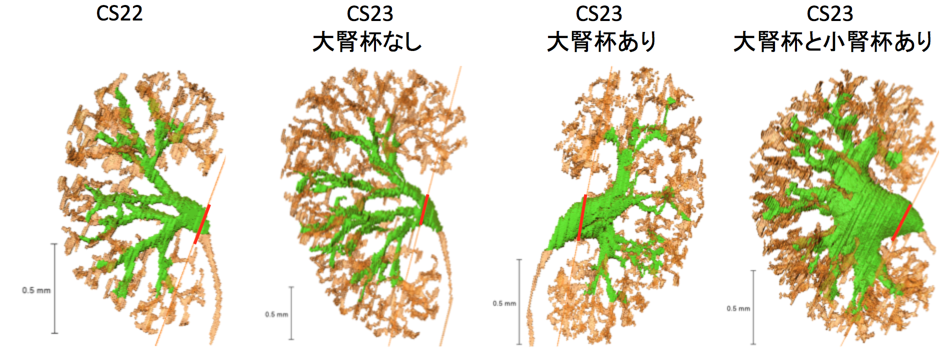
An elaborate system of ducts collects urine from all nephrons, and this structure is known as the urinary collecting system (UCS). This study focused on how the UCS is formed during human embryogenesis. Fifty human embryos between the Carnegie stage (CS) 14 and CS23 were selected from the Kyoto Collection at the Congenital Anomaly Research Center of Kyoto University, Japan. Metanephroses, including the UCS, were segmented on serial digital virtual histological sections. Three-dimensional images were computationally reconstructed for morphological and quantitative analyses. A CS timeline was plotted. It consisted of the 3-D structural morphogenesis of UCS and quantification of the total amount of end-branching, average and maximum numbers of generations, deviation in the metanephros, differentiation of the urothelial epithelium in the renal pelvis, and timing of the rapid expansion of the renal pelvis. The first UCS branching generation occurred by CS16. The average branching generation reached a maximum of 8.74 ± 1.60 and was already the twelfth in CS23. The total end-branching number squared between the start and the end of the embryonic period. UCS would reach the fifteenth branching generation soon after CS23. The number of nephrons per UCS end-branch was low (0.21 ± 0.14 at CS19, 1.34 ± 0.49 at CS23), indicating that the bifid branching occurred rapidly and that the formation of nephrons followed after. The renal pelvis expanded mainly in CS23, which was earlier than that reported in a previous study. The number of nephrons connected to the UCS in the expanded group (246.0 ± 13.2) was significantly larger than that of the pre-expanded group (130.8 ± 80.1) (P < 0.05). The urothelial epithelium differentiated from the zeroth to the third generations at CS23. Differentiation may have continued up until the tenth generation to allow for renal pelvis expansion. The branching speed was not uniform. There were significantly more branching generations in the polar- than in the interpolar regions (P < 0.05). Branching speed reflects the growth orientation required to form the metanephros. Further study will be necessary to understand the renal pelvis expansion mechanism in CS23. Our CS-based timeline enabled us to map UCS formation and predict functional renal capacity after differentiation and growth.
⑦ Miyazaki R, Makishima H, Männer J, Sydow HG, Uwabe C, Takakuwa T, Viebahn C, Yamada S. The Blechschmidt Collection: revisiting specimens from a historical collection of serially sectioned human embryos and fetuses using modern imaging techniques, Congenit Anom, 2018, 58, 152-157, doi: 10.1111/cga.12261
ABSTRACT
Along with the Carnegie Collection in the United States and the Kyoto Collection in Japan, the Blechschmidt Collection (Georg-August-University of Göttingen, Germany) is a major historical human embryo and fetus collection. These collections are of enormous value to human embryology; however, due to the nature of the historical histological specimens, some stains are fading in color, and some glass slides are deteriorating over time. To protect these specimens against such degradation and ensure their future usefulness, we tried to apply modern image scanning and computational reconstruction. Samples of histological specimens of the Blechschmidt Collection were digitized into images using commercial flatbed scanners with a resolution of 4800 pixels per inch. Two specimens were reconstructed into three-dimensional (3D) images by using modern techniques to vertically stack two-dimensional images of the slices into 3D blocks. The larger specimen of crown-rump length (CRL) 64.0 mm, a series of very large histological sections in human embryology, was reconstructed clearly, with its central nervous system segmented before stacking. The smaller specimen of CRL 17.5 mm was also reconstructed into 3D images. The outer surface of the embryo was intact, and its development was classified according to the widely used Carnegie stages (CSs). The CS of the specimen was identified as the later half of CS 20. The invaluable Blechschmidt Collection can be revisited for further research with modern techniques such as digital image scanning and computational 3D reconstruction.
33. Furuichi K, Ishikawa A, Uwabe C, Makishima H, Yamada S, Takakuwa T, Variations of the circle of Willis at the end of the human embryonic period, 2018, 301, 1312-1319, doi:10.1002/ar.23794
ABSTRACT
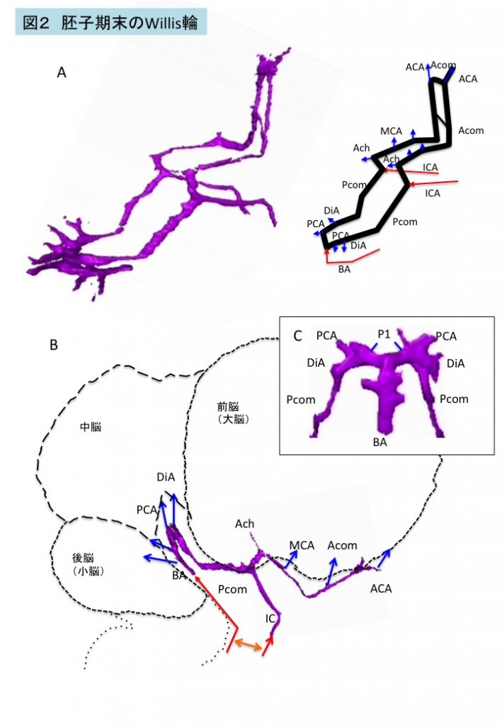
Variations of the circle of Willis (CW) influence blood supply to the brain and adjacent structures in adults. We examined the formation of the CW in 20 human embryo samples at the end of the embryonic period using 3-D reconstructions of serial histological sections. The CW was closed in all samples, and did not form in a single plane, but was composed of multiple stair-like planes. The artery acutely curved at the caudal part of the CW, namely, at the inlet of the basilar artery and bifurcation of the P1 segment of the posterior cerebral artery (PCA), reflecting flexure of the mesencephalon and diencephalon at this stage. Variations were observed in 17 of 20 samples—only anterior parts (anterior communicating artery [Acom] and anterior cerebral artery [ACA]) in 10 samples, only posterior parts (posterior communicating artery [Pcom]) in one sample, and both anterior and posterior parts in six samples. Variations included the Acom formed as partially duplicated in three samples, duplicated in four, plexiform in three, and no channel as a result of a single azygos ACA in one. The ACA formed as duplicated in two, median ACA in two, and right hypoplasia in one. The Pcom formed in hypoplasia of either side in six samples. Variations observed in this study are similar to those observed in fetuses, neonates, and adults. The P1 segment of PCA was very large in all samples. The present observations indicate that variations in the CW are present from the initiation of CW formation.
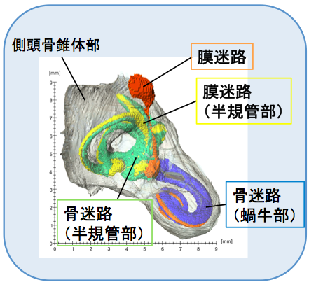
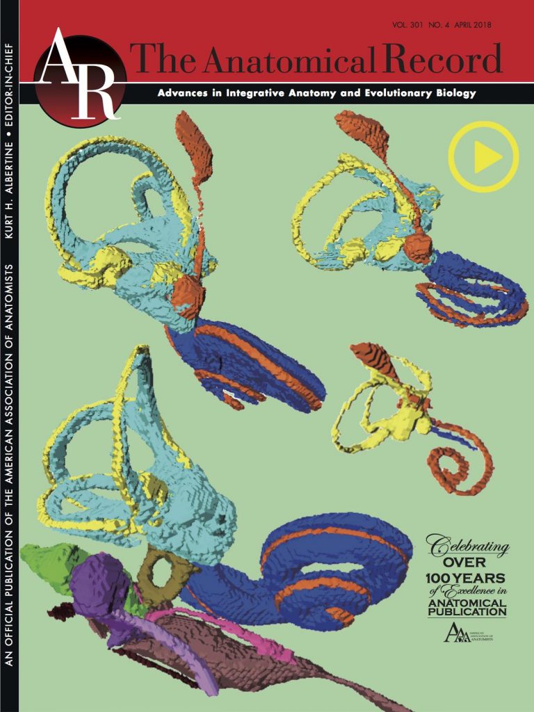
32. Ishikawa A, Ohtsuki S, Yamada S, Uwabe C, Imai H, Matsuda T, Takakuwa T. Formation of the periotic space during the early fetal period in humans, Anat Rec, 2018, 301(4);563-570, 10.1002/ar.23764, 10.1002/ar.23657
Abstract
The inner ear is a very complicated structure, composed of a bony labyrinth (otic capsule; OC), membranous labyrinth, with a space between them, named the periotic labyrinth or periotic space. We investigated how periotic tissue fluid spaces covered the membranous labyrinth three-dimensionally, leading to formation of the periotic labyrinth encapsulated in the OC during human fetal development. Digital data sets from magnetic resonance images and phase-contrast X-ray tomography images of 24 inner ear organs from 24 human fetuses from the Kyoto Collection (fetuses in trimesters 1 and 2; crown—rump length: 14.4–197 mm) were analyzed. The membranous labyrinth was morphologically differentiated in samples at the end of the embryonic period (Carnegie stage 23), and had grown linearly to more than eight times in size during the observation period. The periotic space was first detected at the 35-mm samples, around the vestibule and basal turn of the cochlea, which elongated rapidly to the tip of the cochlea and semicircular ducts, successively, and almost covered the membranous labyrinth at the 115-mm CRL stage or later. In those samples, several ossification centers were detected around the space. This article thus demonstrated that formation of the membranous labyrinth, periotic space (labyrinth), and ossification of the OC occurs successively, according to an intricate timetable.
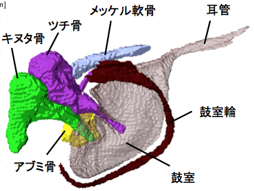
大槻さんの卒論(胎児期の中耳の形成)が The Anatomical Recordに掲載されました。
中耳の耳小骨が骨化する過程、鼓室という空隙で覆われていく様子が立体的に示されました。
CRL 37-197 mm の中耳耳小骨(MEO)の形態形成を検討
MEOの形態は胎児と成人で類似している
CRL150 mm 以降、MEO の大きさはほぼ変わらない
各MEOの骨化は単一の中心から広がる
CRL 86 mmで、鼓室 が明確に観察可能。
31. Ohtsuki S, Ishikawa A, Yamada S, Imai H, Matsuda T, Takakuwa T, Morphogenesis of the middle ear during fetal development as observed via magnetic resonance imaging. Anat Rec 2018, 301, 757-764, doi: 10.1002/ar.23760
ABSTRACT
Recently, our research group has utilized serial histological sections to investigate the morphogenesis of the middle ear, which corresponds to the period of middle ear ossicle (MEO) cartilage formation. However, research regarding middle ear development during the post-embryonic period has been limited. In the present study, we investigated morphogenesis of the middle ear in human fetuses with a crown-rump length (CRL) between 37 and 197 mm using high-resolution magnetic resonance imaging (MRI). Our findings indicated that the morphology of the MEOs is similar during fetal development and adulthood; further, growth of the MEOs nearly ceases once a CRL of 150 mm is attained. In each MEO, ossification spreads from a single center. The malleus and Meckel’s cartilage could be discriminated in samples exhibiting a CRL of 145 mm based on differences in MRI signal intensity. In samples with a CRL of 86 mm, the tympanic cavity (TC) appeared as a thin yet distinct structure attached to the external auditory meatus at the convex surface. Only the handle of the malleus was covered by the TC, while the incus and stapes contacted the cavity at the region of articulation between the two ossicles only, even after a CRL of 145 mm had been attained. Thus, although the TC increased in both diameter and thickness, coverage did not extend across all three MEOs during the observation period. These data are expected to provide a useful standard for morphogenesis and may aid researchers in distinguishing between normal and abnormal development. Anat Rec, 301:757–764, 2018.
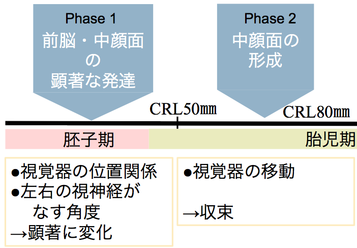
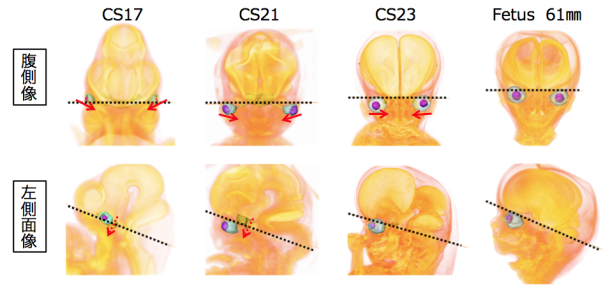
Osaka M, Ishikawa A, Yamada S, Uwabe C, Imai H, Matsuda T, Yoneyama A, Takeda T, Takakuwa T, Positional changes of the ocular organs during craniofacial development, Anatomical Record, 300(12), 2107–2114, 2017 DOI: 10.1002/ar.23588
ABSTRACT
The present study aimed to describe the positional changes of the ocular organs during craniofacial development; moreover, we examined the relationships among the ocular organs and other internal structures. To do this, we traced the positions of the ocular organs in 56 human early fetal samples at different stages of development using high-resolution magnetic resonance imaging and phase-contrast X-ray computed tomography. The eyes were located on the lateral side in the ventral view at Carnegie stage (CS) 16, and then changed their positions medially during development. The eyes remained in the neurocranium until CS17. However, the eyes changed their positions medially and caudally in the viscerocranium after CS18. The positional relationship between the eyes and pituitary gland changed in the lateral view as development progressed. Specifically, they were close to each other at CS17, but moved apart during the later stages of development. These positional changes were also demonstrated quantitatively with morphometric analyses. Based on the present data, the positional changes of the eyes can be categorized into phases, as follows: Phase 1, dramatic positional changes (early fetal period until CS23); and Phase 2, mild positional changes (stabilized; early fetal period after CS23). Notably, all absolute lengths measured in the present study linearly increased as the crown-rump length increased irrespective of the phase, while features of the measured angles and ratios differentially changed in Phases 1 and 2. The present data may help improve our understanding of both the normal and abnormal development of the ocular organs and craniofacial area.
⑭ Katsube M, Yamada S, Miyazaki R, Yamaguchi Y, Makishima H, Takakuwa T, Yamamoto A, Fujii Y, Morimoto N, Ito T, Imai H, Suzuki S, Quantitation of nasal development in the early prenatal period using geometric morphometrics and MRI: A new insight into the critical period of Binder phenotype. Prenatal Diag 37: 907–915, 2017, DOI: 10.1002/pd.5106.2017
Abstract
Objectives
Disturbance of the development of the nasal septum in the early prenatal period causes congenital facial anomalies characterized by a flat nose and defects of the anterior nasal spine (ANS), such as Binder phenotype. The present research aimed to assess the development of the nasal septum and the ANS with growth in the early prenatal period.
Methods
Magnetic resonance images were obtained from 56 specimens. Mid-sagittal images were analyzed by using geometric morphometrics for the development of the nasal septum, and angle analysis was performed for the development of the ANS. Additionally, we calculated and visualized the ontogenetic allometry of the nasal septum.
Results
Our results showed that the nasal septum changed shape in the anteroposterior direction in smaller specimens, while it maintained an almost isometric shape in larger specimens. Furthermore, mathematical evidence revealed that the maturation periods of the shapes of the ANS and the nasal septum were around 12 and 14 weeks of gestation, respectively.
Conclusion
The anteroposterior development of the nasal septum is specific until 14 weeks of gestation, and it is important for nasal protrusion and the development of the ANS. Therefore, the disturbance of such development could induce low nasal deformity, including Binder phenotype.