


食道、胃、十二指腸の前半部は、尾側前腸から形成されます。その境界部すなわち、食道・胃の境界、幽門部の形成について、MRI画像から管全体、管腔それぞれの太さの評価、Tractographyによる筋層線維形成の評価を行い、特徴を明らかにしました。(金橋先生)
- 尾部前腸境界形成の正確なタイムラインをMRI_DTI等を用いて可視化
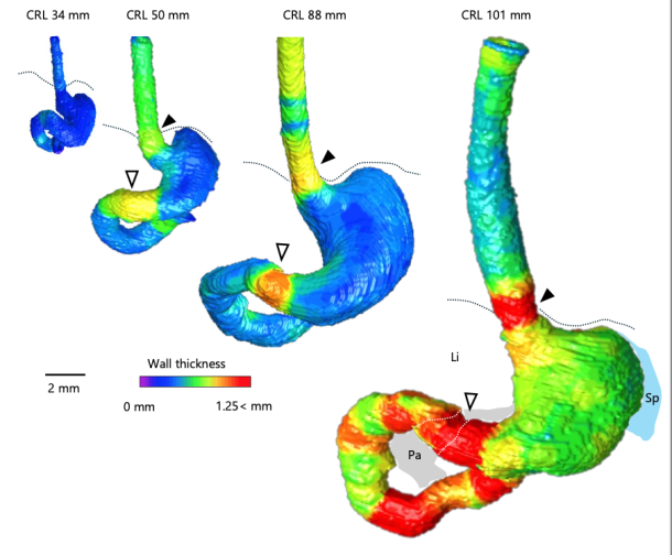
- 食道裂孔および近位幽門における管腔径対壁厚比は、CRL と負の相関関係、壁厚増加は、管腔狭小化をもたらす。
- CRL ≥ 50 mmの個体では、下部食道および幽門管壁が相対的に厚い。
- 下部食道括約筋が位置する食道裂孔の食道壁は、CRL ≥ 88 mmのサンプルで特に厚い。
- 食道裂孔の肥厚した食道壁には厚い粘膜下層があり、幽門管のすべての層が成長とともに肥厚した。幽門管管腔は、遠位部から近位部にかけて狭くなった。
- DTI では、下部食道壁は主に縦線維が検出され、幽門管壁は円形線維が検出される。
- 先天性前腸閉塞の病因に対する理解を深める可能性。
74. KanahashiT, ImaiH, Otani H, YamadaS, Männer J, Takakuwa T. Boundary Formation of the Human Caudal Foregut During the Early Fetal Period: Three-Dimensional Analysis Using T1-Weighted and Diffusion Tensor Images. Cells Tissues Organs, 2025, in press. doi: 10.1159/000546997
Introduction: While caudal foregut development in human fetuses has been outlined in previous research, the formation of its border region remains unclear. This study aimed to visualize the precise timeline of caudal foregut boundary formation. Methods: Three-dimensional images of the foregut from T1-weighted scans of 24 fetuses (crown–rump length [CRL]: 34–103 mm) were analyzed to measure the wall thickness and lumen diameter at nine specific sites. The internal structure in the border region was verified using histological sections and diffusion tensor imaging (DTI) tractography. Results: The lower esophageal and pyloric canal walls in samples with CRL ≥50 mm were relatively thicker. The esophageal wall at the esophageal hiatus, where the lower esophageal sphincter is located, was particularly thick in samples with CRL ≥88 mm. Increased wall thickness at the esophageal hiatus and pyloric canal resulted in a narrower lumen. The pyloric canal lumen narrowed from its distal to proximal sections. The lumen diameter-to-wall thickness ratio at the esophageal hiatus and proximal pyloric was negatively correlated with CRL. The thickened esophageal wall at the esophageal hiatus had a thick submucosa layer, and all layers in the pyloric canal thickened with growth. DTI tractography revealed that the lower esophageal wall mainly comprised longitudinal fibers, whereas the pyloric canal wall consisted solely of circular fibers, with fractional anisotropy increasing with growth. Conclusion: This study provides a comprehensive timeline of normal caudal foregut boundary formation during the early human fetal period, thereby improving the understanding of congenital foregut obstruction pathogenesis.