 「多元計算解剖学」第4回サマーワークショップ(7月14、15日、神戸市)に参加しました。新学術領域研究「医用画像に基づく計算解剖学の多元化と高度知能化診断・治療への展開」研究班のmeetingです。今年度最終年になりました。
|
||||
  |
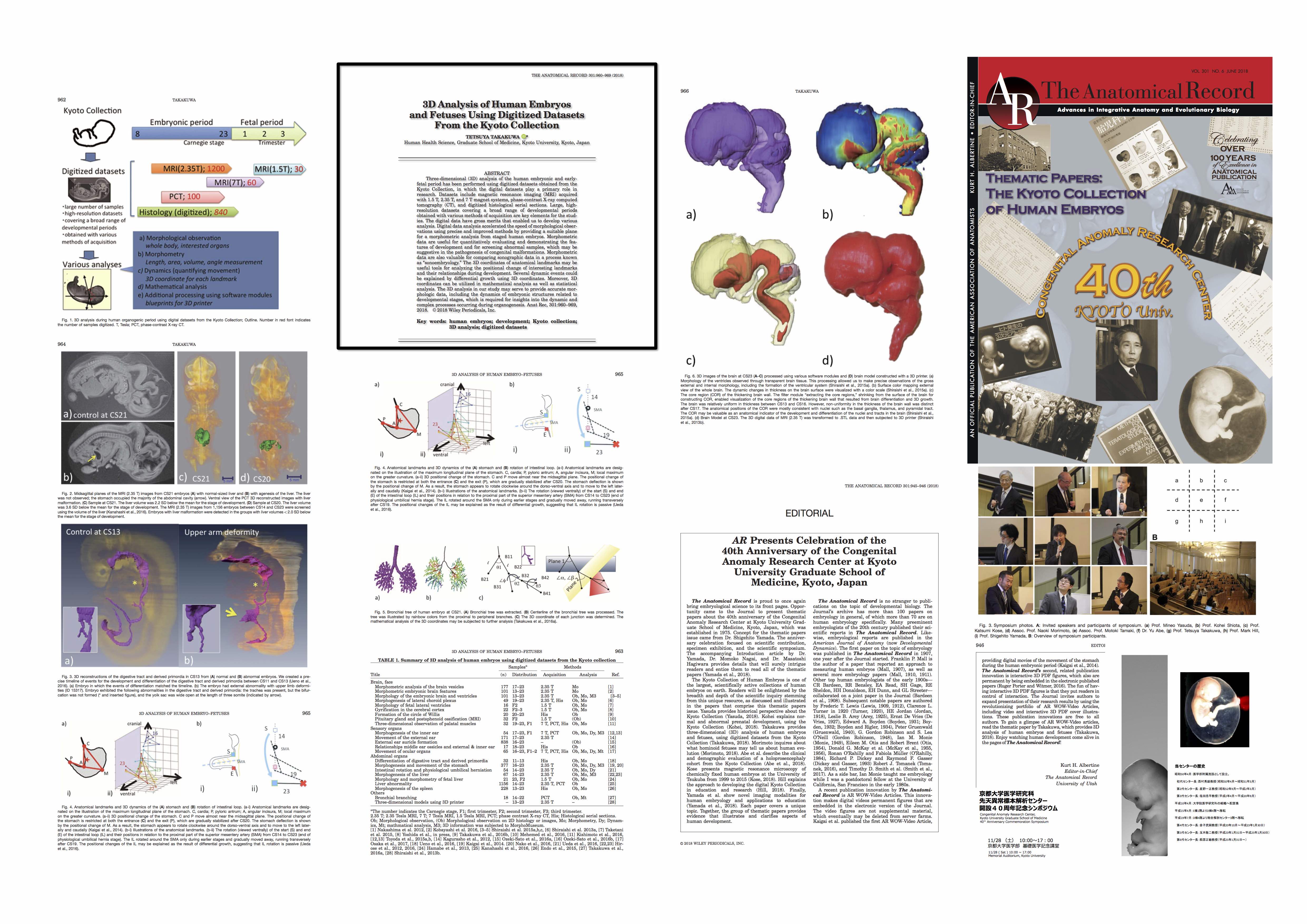
位相CT撮像データからイメージ作成を行う手法を共同研究者の米山先生から習いました。 先天研開設40周年記念号がAnatomical Recに掲載されました。
先天研開設40周年記念号;ヒト胚子・胎児研究についての総説 Anat Rec 2018, 301,960-969 (クリックで拡大されます) 金橋君が2018年度日本先天異常学会奨励賞を受賞しました。おめでとうございます。 Kanahashi T, Yamada S, Tanaka M, Hirose A, Uwabe C, Kose K, Yoneyama A, Takeda T, Takakuwa T, A novel strategy to reveal the latent abnormalities in human embryonic stages from a large embryo collection, Anatomical Record, 299,8-24,2016 10.1002/ar.23281(概要), *299(1),2016の表紙に採用されました。DOI: 10.1002/ar.23206 (cover page) 石川さんの論文がAnatmial Recordに掲載されました。 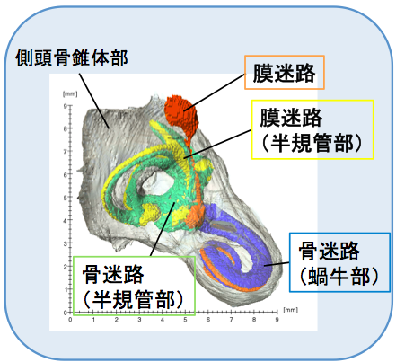 内耳のうち膜迷路、骨迷路の間にあるperiotic spaceの形成に着眼したユニークな論文です。図が表紙に採用されました!!
32. Ishikawa A, Ohtsuki S, Yamada S, Uwabe C, Imai H, Matsuda T, Takakuwa T. Formation of the periotic space during the early fetal period in humans, Anat Rec, 2018, 301(4);563-570, 10.1002/ar.23764, 10.1002/ar.23657 Abstract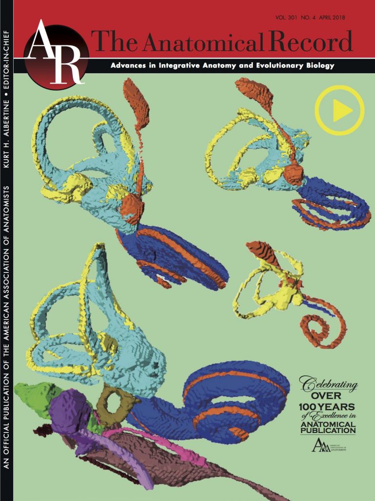 The inner ear is a very complicated structure, composed of a bony labyrinth (otic capsule; OC), membranous labyrinth, with a space between them, named the periotic labyrinth or periotic space. We investigated how periotic tissue fluid spaces covered the membranous labyrinth three-dimensionally, leading to formation of the periotic labyrinth encapsulated in the OC during human fetal development. Digital data sets from magnetic resonance images and phase-contrast X-ray tomography images of 24 inner ear organs from 24 human fetuses from the Kyoto Collection (fetuses in trimesters 1 and 2; crown—rump length: 14.4–197 mm) were analyzed. The membranous labyrinth was morphologically differentiated in samples at the end of the embryonic period (Carnegie stage 23), and had grown linearly to more than eight times in size during the observation period. The periotic space was first detected at the 35-mm samples, around the vestibule and basal turn of the cochlea, which elongated rapidly to the tip of the cochlea and semicircular ducts, successively, and almost covered the membranous labyrinth at the 115-mm CRL stage or later. In those samples, several ossification centers were detected around the space. This article thus demonstrated that formation of the membranous labyrinth, periotic space (labyrinth), and ossification of the OC occurs successively, according to an intricate timetable. 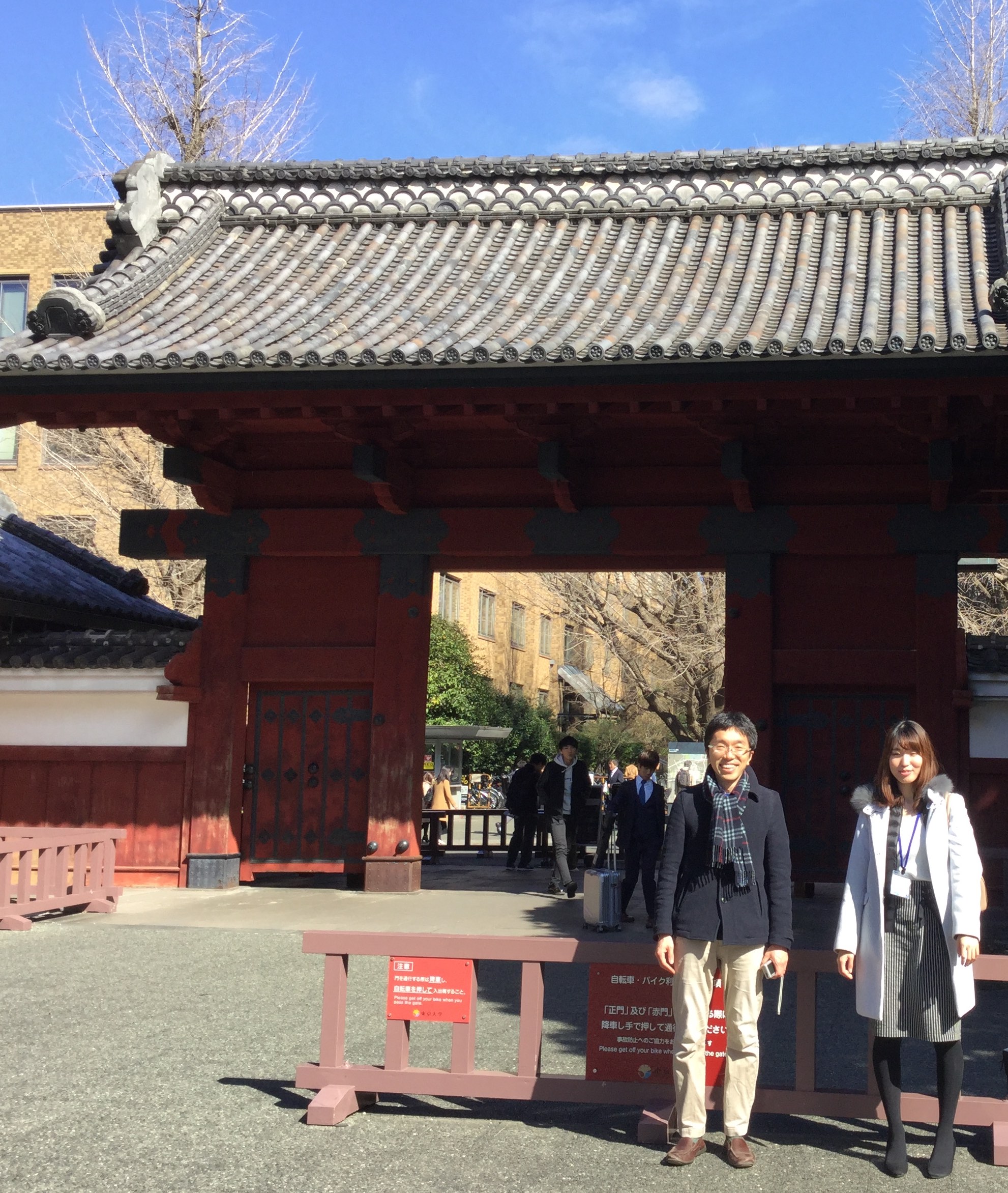 赤門前, 共同研究者;松林先生、藤井さん 多元計算解剖学第4回国際シンポジウム(The 4th International Symposium on Multidisciplinary Computational Anatomy)で発表しました。(3/2-2/3, 東京大学) A02-KB107 Analysis of Central Nervous System and Skeletal System During Human Early-fetal Period Based on Multidisciplinary Computational Anatomy -Progress Overview FY 2017- (PI:Tetsuya Takakuwa) 2017年度; 修士論文発表会が行われました(2018.0207; 杉浦ホール) ヒト胚子期における腎盂形成の三次元的解析 石山 華 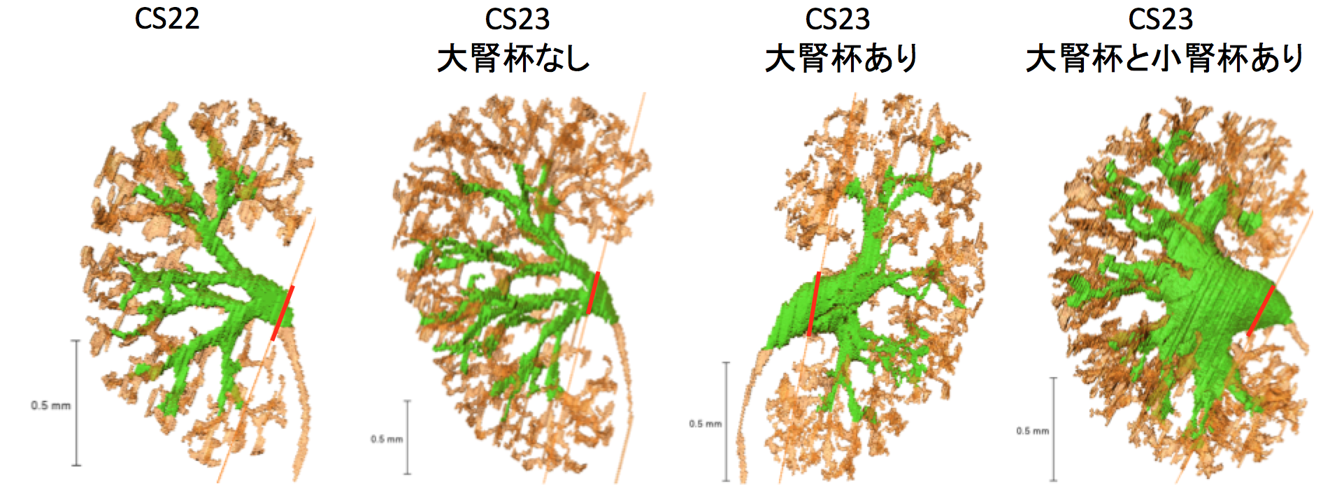 【背景】腎盂の発生はCarnegie Stage(CS)14頃に始まる。この発生過程については、組織切片を用いた平面的な研究が主であり、CSに沿った報告としてShikinamiの報告がある。 37. Ishiyama H, Ishikawa A, Imai H, Matsuda T, Yoneyama A, Yamada S, Takakuwa T. Spatial relationship between the metanephros and adjacent organs according to the Carnegie stage of development. Anat Rec (Hoboken) 2019. 302, 1887-2104. DOI: 10.1002/ar.24103 34. Ishiyama H, Ishikawa A, Kitazawa H, Fujii S, Matsubayashi J, Yamada S, Takakuwa T, Branching morphogenesis of the urinary collecting system in the human embryonic metanephros, PLoS One 13(9): e0203623. doi: 10.1371/journal.pone.0203623 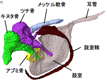 大槻さんの卒論(胎児期の中耳の形成)が The Anatomical Recordに掲載されました。 中耳の耳小骨が骨化する過程、鼓室という空隙で覆われていく様子が立体的に示されました。
31. Ohtsuki S, Ishikawa A, Yamada S, Imai H, Matsuda T, Takakuwa T, Morphogenesis of the middle ear during fetal development as observed via magnetic resonance imaging. Anat Rec 2018, 301, 757-764, doi: 10.1002/ar.23760 ABSTRACTRecently, our research group has utilized serial histological sections to investigate the morphogenesis of the middle ear, which corresponds to the period of middle ear ossicle (MEO) cartilage formation. However, research regarding middle ear development during the post-embryonic period has been limited. In the present study, we investigated morphogenesis of the middle ear in human fetuses with a crown-rump length (CRL) between 37 and 197 mm using high-resolution magnetic resonance imaging (MRI). Our findings indicated that the morphology of the MEOs is similar during fetal development and adulthood; further, growth of the MEOs nearly ceases once a CRL of 150 mm is attained. In each MEO, ossification spreads from a single center. The malleus and Meckel’s cartilage could be discriminated in samples exhibiting a CRL of 145 mm based on differences in MRI signal intensity. In samples with a CRL of 86 mm, the tympanic cavity (TC) appeared as a thin yet distinct structure attached to the external auditory meatus at the convex surface. Only the handle of the malleus was covered by the TC, while the incus and stapes contacted the cavity at the region of articulation between the two ossicles only, even after a CRL of 145 mm had been attained. Thus, although the TC increased in both diameter and thickness, coverage did not extend across all three MEOs during the observation period. These data are expected to provide a useful standard for morphogenesis and may aid researchers in distinguishing between normal and abnormal development. Anat Rec, 301:757–764, 2018. |
|||