気管支分岐の法則性について
新学術領域研究「多元計算解剖学」第2回 国際シンポジウムで発表しました。
Three-dimensional Analysis of the Bronchial Branching in Human Embryonic Stages
Takakuwa T, Muranaka T, Yamada S
日時:平成28年2月11日 (木・祝)9:00~18:00
2月12日 (金) 9:00~16:50
場所:名古屋大学 野依記念学術交流館 2階
|
||||
  |
気管支分岐の法則性について 新学術領域研究「多元計算解剖学」第2回 国際シンポジウムで発表しました。 Three-dimensional Analysis of the Bronchial Branching in Human Embryonic Stages Takakuwa T, Muranaka T, Yamada S
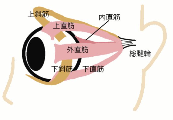
日時:平成28年2月11日 (木・祝)9:00~18:00 2月12日 (金) 9:00~16:50 場所:名古屋大学 野依記念学術交流館 2階 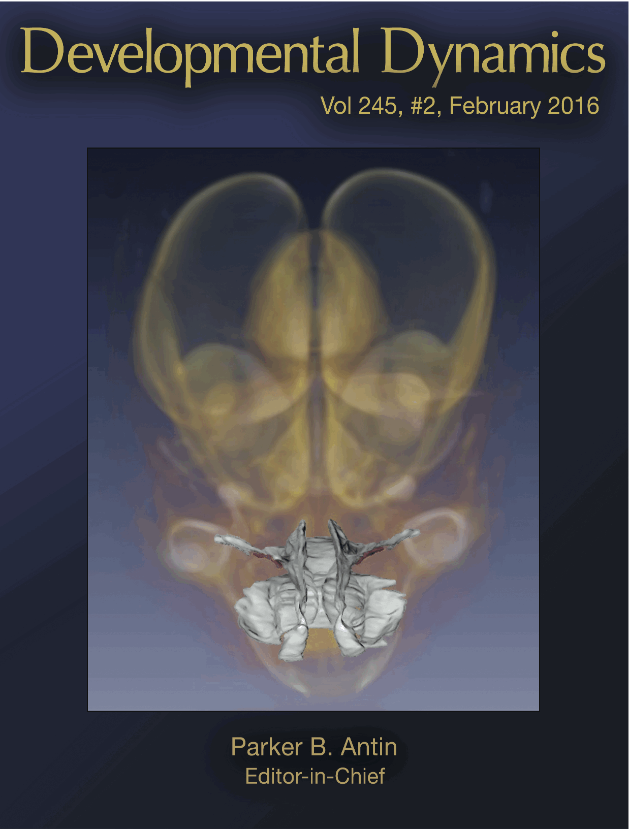 金橋君の描いた図がDevelopmental Dynamicsの表紙に採用されました。 岸本先生(先天異常標本解析センター)の研究の一部として、Amiraを用いてヒト胎児の咽頭口蓋領域の解析の補助を金橋くん(修士)が行いました。 ① Kishimoto H, Yamada S, Kanahashi T, Yoneyama A, Imai H, Matsuda T, Takeda T, Kawai K, Three-dimensional observation of palatal muscles in the human embryo and fetus: development of levator veli palatini and clinical importance of the lesser palatine nerve, Developmental Dynamics 245: 123–131, 2016, DOI: 10.1002/dvdy.24382, DOI: 10.1002/dvdy.24364 植田さんの卒業研究が、Anatomical Recに受諾されました。 ヒト胚子期の中腸の発生過程でみられる、腸の回転と臍帯内への生理的ヘルニアについての定量的な研究です。 
19. Ueda Y, Yamada S, Uwabe C, Kose K, Takakuwa T, Intestinal rotation and physiological umbilical herniation during the embryonic period, Anatomical Record 299, 197-206, 2016, DOI: 10.1002/ar.23296 ABSTRACTDrastic changes occur during the formation of the intestinal loop (IL), including elongation, physiological umbilical herniation (PUH), and midgut rotation. Fifty-four sets of magnetic resonance images of embryos between Carnegie stage (CS) 14 and CS 23 were used to reconstruct embryonic digestive tract in three dimensions in the Amira program. Elongation, PUH, and rotation were quantified in relation to the proximal part of the superior mesenteric artery (SMA), designated as the origin. Up to CS 16, IL rotation was initially observed as a slight deviation of the duodenum and colorectum from the median plane. The PUH was noticeable after CS 17. At CS 18, the IL showed a hairpin-like structure, with the SMA running parallel to the straight part and the cecum located to the left. After CS 19, the IL began to form a complex structure as a result of the rapid growth of the small intestinal portion. By CS 20, the IL starting point had moved from the right cranial region to an area caudal to the origin, though elongation of the duodenum was not conspicuous—this was a change of almost 180° in position. The end of the IL remained in roughly the same place, to the left of and caudal to the origin. Notably, the IL rotated around the origin only during earlier stages and gradually moved away, running transversely after CS 19. The movements of the IL may be explained as the result of differential growth, suggesting that IL rotation is passive. ヒト器官形成期における視覚器の発達についての3次元的解析 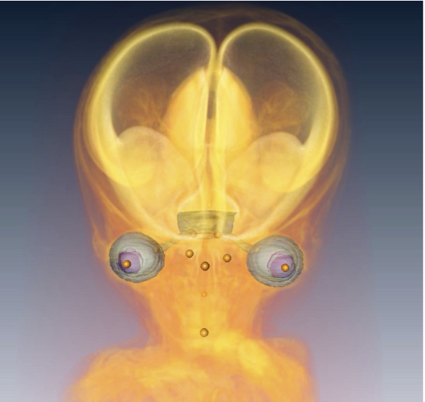 【背景】視覚器の発生はCarnegie Stage(CS) 10の視溝形成から始まり、生後数ヶ月まで発達が続く。器官形成期における組織学的な研究はこれまで多くの報告があるが、3次元的な検討は十分には行われていない。 【対象と方法】京都大学大学院医学研究科附属先天異常標本解析センター所有のヒト胚子、胎児標本の立体情報計65例(位相CTdata;26例、MRIdata;39例)を対象とした。用いた標本は全て明らかな外表奇形、視覚器の異常を伴っておらず、胚子標本はCS16~CS23、胎児標本は胎児期初期(CRL34㎜~152㎜)に分類される。画像情報を基に視覚器の立体像を作製し、1)頭部における水晶体や網膜(強膜)、視神経などの立体的位置関係の変化の検討、2)外眼筋の形成過程の検討を行った。 【結果】1)視覚器の立体的位置関係の変化: 水晶体は外側から腹側、頭側から尾側へ移動した。頭部横径に対する水晶体中心間距離はCS16~C21では1~1.2だったが成長に伴い減少し、CS16~CRL30㎜の胎児期初期までは変化が大きく、その後0.4~0.5に収束した。頭尾方向の移動はCS17~CS18 にかけて大きな変化がみられた。左右の視神経がなす角度は、CS16~17では160°~180°だったが徐々に小さくなりCRL30㎜の胎児期以降では60°~80°に収束した。いずれの変化もCRL30㎜以降の胎児期初期で収束し成人の値と近くなることから、この時期から頭部における視覚器の位置関係は成人に近いことを示した。 2)外眼筋の形成過程: 胎児期初期では成人と同様、4本の直筋(上直、下直、内直、外直)と上斜筋は視神経管周囲の骨膜(総腱輪)から、下斜筋は眼窩の前縁内側骨膜から起始していた。全ての外眼筋が眼球表面の強膜に停止しており、発達に大きな左右差はみられなかった。体積は外直筋、長さは上斜筋、平均断面積は上直筋が最も増加率が大きかった。上斜筋の滑車部分がなす角度は、CS22~23では直角に近かったが、徐々に小さくなりCRL50㎜以降の胎児期では40°~50°に収束し成人と近い値を示した。 【結論】胚子期、胎児期初期のいずれも正常と判定された個体の位相CTdataやMRIdataを用いて視覚器の立体像を作成し、成長に伴う視覚器の立体的位置関係の変化や外眼筋の成長を定量的に明らかにした。異常個体の立体像も同様に作成することで、視覚器の発生の異常についても解析できることが期待できる。また、さらに解像度の高い撮像方法が可能となることで、今回解析できなかったCS16以降の個体でも詳細な解析が行われることが期待できる。 29. Osaka M, Ishikawa A, Yamada S, Uwabe C, Imai H, Matsuda T, Yoneyama A, Takeda T, Takakuwa T, Positional changes of the ocular organs during craniofacial development, Anat Rec (Hoboken) 300(12), 2107–2114, 2017 DOI: 10.1002/ar.23588(概要) 4回生の卒業研究発表会が行われました。 本年度は、理工系のグループに混じっての発表になりました。 ヒト胚子期~胎児期における側頭骨錐体部の内部構造の三次元的観察 石川 葵 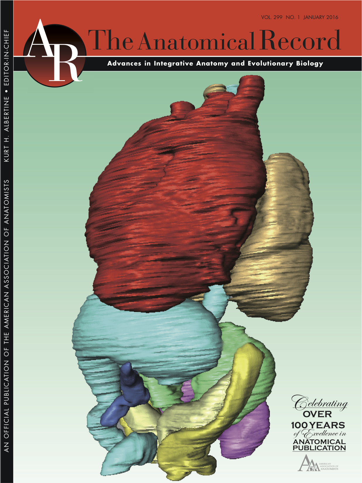 金橋くんの論文の図がAnatomical Record 299巻1号の表紙に採用されました。位相CTを用いて解析した肝臓のない個体です。同個体は胎児期に発生が進まなくなり流産すると考えられます。 金橋くんの論文は同号に掲載されました。 18. Kanahashi T, Yamada S, Tanaka M, Hirose A, Uwabe C, Kose K, Yoneyama A, Takeda T, Takakuwa T, A novel strategy to reveal the latent abnormalities in human embryonic stages from a large embryo collection, Anatomical Record, 299,8-24,2016 10.1002/ar.23281(概要), DOI: 10.1002/ar.23206 (cover page) 京都大学医学研究科先天異常標本解析センター 開設40周年記念シンポジウムが開催されました。 日時 平成27年 11月 28日(土)10時~17時 場 所 京都大学 基礎医学記念講堂 講演者 安田峯生 広島大学 名誉教授 主 催 京都大学医学研究科 先天異常標本解析センター *高桑も研究室の研究内容を発表いたしました。 |
|||