Marieさんが大学院教育支援機構奨励研究員及びフェローシップ受給者によるポスター発表会・研究交流会で発表しました(2022.10.21)
Saizonou Marie Ange ; Understanding the development and differentiation of epithelium of Urinary Collecting System in human embryonic metanephros
|
||||
  |
Marieさんが大学院教育支援機構奨励研究員及びフェローシップ受給者によるポスター発表会・研究交流会で発表しました(2022.10.21) Saizonou Marie Ange ; Understanding the development and differentiation of epithelium of Urinary Collecting System in human embryonic metanephros  掛谷さんの修士論文がJ Anatomyに掲載されました。 発生途中、消化管は臍帯腔内に脱出し、CRL40mmころに短時間で還納されます。還納の仕方は、これまで消化管の動きを中心に研究されており、slide-stack model(消化管はループを形成したまま還納するというモデル)が優勢でした。本論文では、下記の2つの定説を覆す内容です。
複雑な小腸の走行を追跡しても限界があることを認識し、この論文では消化管を栄養する上腸間膜動脈とその小腸枝の走行を正確に追うことで、還納経過中の小腸の位置や形状、走行をしめすとともに、血管系の分布、形状変化も示すことができました。還納は腸管の動きとして認識される時期に先行する血管系の位置の変化により開始され、これまでのコンセンサスよりも早く始まることを示しました。臍帯輪通過時には、消化管とそれを栄養する動脈の走行は形状を変えます。消化管はループがほどけ、臍帯輪を2本以上の消化管が通過することはありません。また、臍帯輪において消化管、腸間膜、動脈は整然とならび、どの個体でもほぼ一定です。 この組織だった配置は腸管への血行が安全に確保されるために必要とかんがえられます。 KakeyaM, Matsubayashi J, Kanahashi T, Männer J, Yamada S, Takakuwa T. The return process of physiological umbilical herniation in human fetuses: the possible role of the vascular tree and umbilical ring. J Anatomy 2022, 241(3), 846-859. https://doi.org/10.1111/joa.13720 AbstractThe human intestine elongates during the early fetal period, herniates into the extraembryonic coelom (EC), and subsequently returns to the abdominal cavity (AC). The process by which the intestinal loop returns to the abdomen remains unclear. This study aimed to document positional changes in the intestinal tract with the superior mesenteric artery (SMA) and branches in 3D to elucidate the intestinal loop return process (transition phase). Serial histological cross-sections from human fetuses (crown–rump length [CRL] range: 30–50 mm) in the herniation (n = 1), transition (n = 7), and return (n = 2) phases were selected from the Blechschmidt Collection. The distribution of the SMA trunk and all intestinal and sister branches entering the intestines was visualized so that positional changes in branches were continuous from the herniation to return phases. Positional changes in SMA branches proceeded in an orderly and structured manner; this is essential for continuous blood supply via the SMA to the intestine during transition and for safe intestinal return. Changes in the SMA distribution proceeded prior to the detection of initiation of intestinal tract return, which might start earlier and last much longer than our consensus (i.e., that the return of the herniated intestine begins when the CRL is approximately 40 mm and ends within a short time). In the cross-section of the umbilical ring in the herniation and transition phases, one proximal limb and one distal limb were observed with SMA intestinal branches, which were fully packed in the umbilical ring. The SMA branches were aligned from inferior to superior along the SMA main trunk. In the herniation phase, the distribution of 3rd–13th branches aligned from proximal inferior medial to distal superior left with a slight spiral in the EC, the tips of which suggested an orderly running course of the small intestine. In the transition phase, SMA branches running across the umbilical ring that fed the small intestine were observed, suggesting that the intestine was uncoiled and ran across the umbilical ring almost vertically. The estimated curvature value supported the phenomenon of uncoiling at the umbilical ring; the value at the umbilical ring was lesser than that in the AC and EC. During the transition phase, the proximal and distal limbs transversely ran side by side in the AC, umbilical ring, limbs on the cranial side, and mesentery on the caudal side. The SMA trunk and its branches ran in parallel, cranially to caudally aligned in the mesentery. This layout of the umbilical ring was maintained during the transition phase. In the return phase, the SMA trunk was gently curved from the upper left to the lower right of the AC; around 12 branches spread with a winding staircase appearance. The intestinal tract reached its definitive position immediately after all tissues crossed the umbilical ring and released any restriction. Each SMA branch and the corresponding region of the intestinal tract form a unit and change their position, though the conformation may change within each unit when running across the umbilical ring. We suggest that the slide–stack model requires revision. 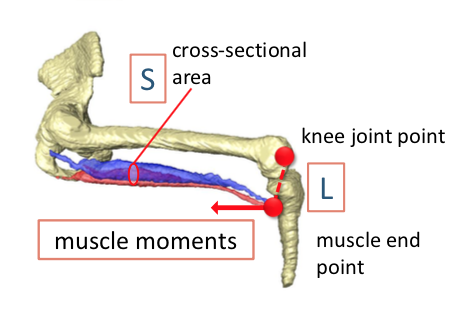 The 20th Congress of International Federation of Associations of Anatomists (IFAA2022) 2022,8/5-7, Istanbul,Turkey/Onlineで発表しました。 Ishikawa A, Nagai-Tanima M, Ishida K, Imai H, Aoyama T, Takakuwa T. Three-dimensional analysis of knee joint development during the human fetal period.  第62回日本先天異常学会で発表しました(‘22,7/29-31, 金沢) 酷暑の中、対面、遠隔のハイブリッドで開催されました。 シンポジウム(正常と異常を顕かにするイメージング技術) 高桑徹也;三次元デジタル情報を活用したヒト胚子・胎児の解析 @座長もさせていただきました。現地に出席の方も結構おられ、かなり盛況でした。 一般演題 藤井 瀬菜、村中 太河、松林 潤、米山 明男、兵藤 一行、山田 重人、高桑徹也; ヒト胚子期における気管支樹の三次元的変化の定量的検討 金橋 徹、今井宏彦、大谷 浩、山田重人、米山明男、高桑徹也; 拡散テンソルイメージングを応用したヒト胎児横隔膜形成の三次元的解析 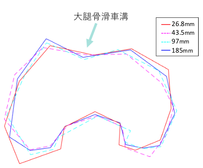 JOSKAS-JOSSM 2022 第14回日本関節鏡・膝・スポーツ整形外科学会/第48回日本整形外科スポーツ医学会学術集会で発表しました。(札幌コンベンションセンター、北海道、2022,6/16〜18) 石川 葵、谷間 桃子、石田 かのん、青山 朋樹「ヒト胎児期における膝関節発生の三次元的解析」 石田 かのん、谷間 桃子、石川 葵、青山 朋樹「ヒト胎児期における膝関節発生の三次元的解析」 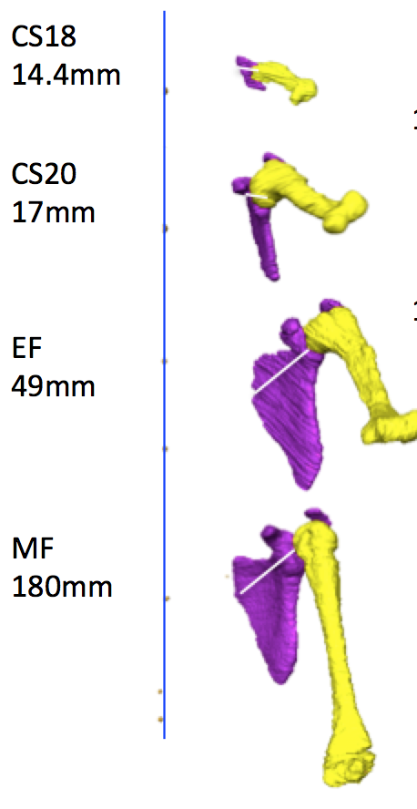 熊野くんの卒業研究のうち上肢の解析分がAnatomical Recに掲載されました。 胚子期後期から胎児期初期、中期についての上肢の肢位について、上腕骨と体幹、体幹と肩甲骨、肩甲骨と上腕と解剖学的にわけて検討しています。
Kumano Y, Tanaka S, Sakamoto R, Kanahashi T, Imai H, Yoneyama A, Yamada S, Takakuwa T. Upper arm posture during human embryonic and fetal development. Anatomical Rec 2022, 305 1682-1691, https://doi.org/10.1002/ar.24796 AbstractThe upper extremity posture is characteristic of each Carnegie stage (CS), particularly between CS18 and CS23. Morphogenesis of the shoulder joint complex largely contributes to posture, although the exact position of the shoulder joints has not been described. In the present study, the position of the upper arm was first quantitatively measured, and the contribution of the position of the shoulder girdle, including the scapula and glenohumeral (GH) joint, was then evaluated. Twenty-nine human fetal specimens from the Kyoto Collection were used in this study. The morphogenesis and three-dimensional position of the shoulder girdle and humerus were analyzed using phase-contrast X-ray computed tomography and magnetic resonance imaging. Both abduction and flexion of the upper arm displayed a local maximum at CS20. Abduction gradually decreased until the middle fetal period, which was a prominent feature. Flexion was less than 90° at the local maximum, which was discrepant between appearance and measurement value in our study. The scapular body exhibited a unique position, being oriented internally and in the upward direction, with the glenoid cavity oriented cranially and ventrally. However, this unique scapular position had little effect on the upper arm posture because the angle of the scapula on the thorax was canceled as the angle of the GH joint had changed to a mirror image of that angle. Our present study suggested that measuring the angle of the scapula on the thorax and that of the GH joint using sonography leads to improved staging of the human embryo.  Early fetal period DTI atlas (画面をクリック) 研究室で進めている胎児期初期の解析をまとめたサイトを現在立ち上げ中です。高解像度MRI_T1強調像を基盤にDTI画像、立体再構成像等の解析結果を順次発表してまいります。ご期待ください。 藤井さん、村中くんの研究がJ Anatomyの’top cited article (2020-21)’に選ばれました。多くの研究者に読んで、評価していただいた結果と思います。おめでとうございます。
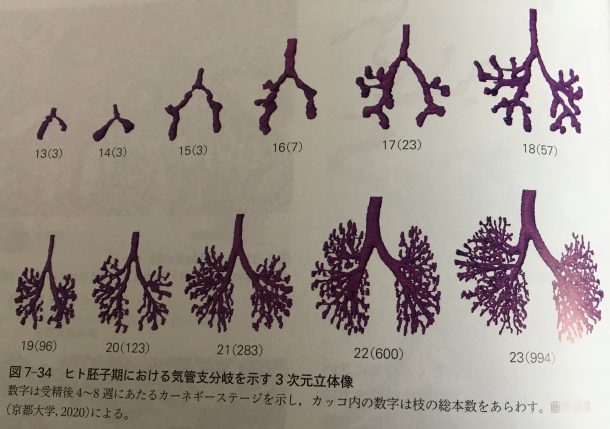
44. Fujii S, Muranaka T, Matsubayash J, Yamada S, Yoneyama A, Takakuwa T. The bronchial tree of the human embryo: an analysis of variations in the bronchial segments. J Anatomy 2020, 237, 311-322. doi: 10.1111/joa.13199. 胚子の気管支の形成過程についての立体再構成像は、標準組織学(医学書院)の図に引用されました。
|
|||