


神楽所さんの卒論「外耳の動きをdifferential growthで説明する」がHead Face Medicineに掲載されました。
『ヒト胚子期に外耳は顔の側方を頭側に大きく移動する』と、多くの発生学の教科書に記載されている。 この動きは”移動 (migration)”ではなく ”分化・成長 (differential growth)” で説明できることを、われわれ は、MRIデータを用いて、個体の中心に基準点をおき、位置変化の絶対値を検討することで明らかにした。 実際の観察では、目や口といった顔の表面にある解剖学的基準をもとに相対的な動きとしてとらえため、外耳は移動してみえるのである。Head Face Med. 2012 Feb 1;8(1):2.
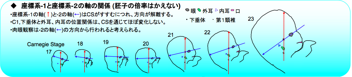
- 外耳の動きが成長差によって説明できるかどうかを検討
- 動きの評価のために、2 つの異なる基準軸を選択
- 下垂体とC1を基準軸; 外耳は主に横方向に移動し、頭側には動かない
- 表面ランドマーク(眼と口を基準軸); 外耳は尾側外側腹領域から眼、口の間に移動
- 外耳が眼や口などの顔の他のランドマークとの相対的な位置関係がある場合、外耳は頭側で動いているように見える
- 結果は、外耳と内耳を含むすべての解剖学的ランドマークの動きが、Differential growthによって説明可能なことを示す
")
Abstract
External ears, one of the major face components, show an interesting movement during craniofacial morphogenesis in human embryo. The present study was performed to see if movement of the external ears in a human embryo could be explained by differential growth. In all, 171 samples between Carnegie stage (CS) 17 and CS 23 were selected from MR image datasets of human embryos obtained from the Kyoto Collection of Human Embryos. The three-dimensional absolute position of 13 representative anatomical landmarks, including external and internal ears, from MRI data was traced to evaluate the movement between the different stages with identical magnification. Two different sets of reference axes were selected for evaluation and comparison of the movements. When the pituitary gland and the first cervical vertebra were selected as a reference axis, the 13 anatomical landmarks of the face spread out within the same region as the embryo enlarged and changed shape. The external ear did move mainly laterally, but not cranially. The distance between the external and internal ear stayed approximately constant. Three-dimensionally, the external ear located in the caudal ventral parts of the internal ear in CS 17, moved mainly laterally until CS 23. When surface landmarks eyes and mouth were selected as a reference axis, external ears moved from the caudal lateral ventral region to the position between eyes and mouth during development. The results indicate that movement of all anatomical landmarks, including external and internal ears, can be explained by differential growth. Also, when the external ear is recognized as one of the facial landmarks and having a relative position to other landmarks such as the eyes and mouth, the external ears seem to move cranially.