藤岡さんの修士論文「軟骨最表層の構造について」がOsteoarthritis Cartilageに掲載されました。
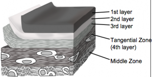
- 関節腔に直接面する最表面ゾーン (MSZ) の構造と分子成分をブタ 膝で組織学的に検討
- MSZ が 3 つの層に細分
- MSZ の最内側 (3 番目) の層;Collagen subtype I, II, III が存在
- tangential layer;3 番目の層の下にあり、type II collagenと少量type III collagenが存在
7. Fujioka R, Aoyama T, Takakuwa T, The layered structure of the articular surface, Osteoarthritis Cartilage, 2013, 21, 1092-1098 doi: 10.1016/j.joca.2013.04.021
■ 内容を詳しく>>
Summary
Objective
Articular cartilage is roughly separated into three areas: the tangential, middle, and deep zones. The structure and molecular components of an additional important zone, the most superficial zone (MSZ), which directly faces the joint cavity, have yet to be conclusively elucidated. The purpose of the present study was to use multiple methods to study the MSZ in order to determine its structure.
Materials and methods
Knees from 16 pigs (age, 6 months) were used. Full-thickness cartilage specimens were harvested from the femoral groove. The MSZ was observed using light microscopy, transmission electron microscopy (TEM), and scanning electron microscopy (SEM) in combination with histochemical and immunohistochemical methods.
Results
The combined findings from the three different observational methods indicate that the MSZ is subdivided into three layers. Among these three layers, collagen subtypes I, II, and III are present in the innermost (third) layer of the MSZ. Beneath the third layer, type II collagen is the predominant type, with small amounts of type III collagen. This layer beneath the third layer is considered to be the tangential layer.
Conclusions
Our observations indicate that the MSZ is subdivided into three layers. Further analysis of the molecular components in each layer may improve our understanding of the structure of the articular surface.


