アクセスカウンター

2016年9月
 1234
567891011
12131415161718
19202122232425
2627282930  

尾関さんの修論がAnat Recに掲載

CS22聴覚器

尾関さんの修士論文のうち、中耳耳小骨の形成と外耳、内耳との立体的位置関係についてAnat Recに掲載されました。

MEO の形成と外耳と内耳の接続のタイムラインを決定しました。

  • 軟骨頭蓋は、CS18 までに認識可能
  • 軟骨形成開始は耳小骨(MEO)間で若干の相違。
  • CS19;槌骨、キヌタ骨、アブミ骨の順序で、前後方向に配置 CS22以降;MEO同士が関節を介し接続。
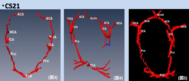
  • CS21以降にすべてのMEOの軟骨形成を確認
  • CS23;アブミ骨基底部分はfoot plate状ではない
  • ツチ骨のハンドルは 外耳道からは離れている
  • 耳介軟骨と耳嚢の軟骨膜境界は明確

26. Ozeki-Satoh M, Ishikawa A, Yamada S, Uwabe C, Takakuwa T. Morphogenesis of the Middle Ear Ossicles and Spatial Relationships with the External and Inner Ears during the Embryonic Period, Anat Rec 299:1325–1337, 2016, DOI 10.1002/ar.23457

Abstract

We describe the three-dimensional morphogenesis of the middle ear ossicles (MEOs) according to Carnegie stage (CS) in human embryos. Seventeen samples including 33 MEOs from CS18 to 23 were selected from the Kyoto Collection. The primordia of the MEOs and related structures were histologically observed and three-dimensionally reconstructed from digital images. The timing of chondrogenesis was variable among structures. The stapes was recognizable as a vague condensation of the mesenchymal cells in all samples from CS18, whereas the malleus and incus were recognizable at CS19. Chondrogenesis of all MEOs was evident in all samples after CS21. The chondrocranium was recognizable in all samples by CS18, and the perichondrium border of the auricular cartilage and otic capsule was distinct in all samples at CS23. At CS19, the MEOs were positioned in the anterior to posterior direction, following the order malleus, incus, stapes, which adjusted gradually during development. The MEOs connected in all samples after CS22. The stapes was located close to the vestibular part of the inner ear, although the basal part was not differentiated into the “footplate” form, even at CS23. The handles of the malleus were close to the tubotympanic recess at CS23, but were distant from the external auditory meatus. Determining the timeline of the formation of MEOs and connection of the external and inner ears can be informative for understanding hearing loss caused by failure of this connection. These data may provide a useful standard for morphogenesis, and will contribute to distinguishing between normal and abnormal MEO development. 

2016次世代医療を語る -再生医療の実用化に向けて-

今年度の研究科横断型プログラム、次世代医療を語る -再生医療の実用化に向けて-(後期水曜5限)の概要が決定しました。

※ 京都大学オープンコースウェアに収録済みです。御覧ください。

OCW_2016次世代医療を語る -再生医療の実用化に向けて(外部)

開講時間:16時30分~18時(5限目)
開講場所:杉浦地域医療センター

【カテゴリーA:再生医学の基礎】
第一回 (10月5日) 幹細胞とは?
中川 誠人(京都大学iPS細胞研究所 未来生命科学開拓部門:講師)

第二回 (10月12日) ヒトiPS細胞の樹立とその有用性
浅香 勲(京都大学iPS細胞研究所 基盤技術研究部門:教授)

第三回 (10月19日)  神経堤細胞と運動器再生
池谷 真(京都大学iPS細胞研究所 未来生命科学開拓部門:准教授)

【カテゴリーB:再生医療の臨床応用】
第四回 (10月26日)   再生医療の足場材料の開発
山本 雅哉(京都大学再生医科学研究所 生体材料学分野:准教授)

第五回 (11月2日)  心臓再生
山下 潤(京都大学iPS細胞研究所 増殖分化機構研究部門:教授)

第六回 (11月9日) 骨格筋再生医療の実現化に向けて
櫻井 英俊 (京都大学iPS細胞研究所 臨床応用研究部門:准教授)

【カテゴリーC:再生医療の実施】
第七回 (11月16日) 再生医療における倫理
八代 嘉美(京都大学iPS細胞研究所 上廣倫理研究部門:准教授)

第八回 (11月30日) 再生医療にかかる規制
伊藤 達也 (京都大学医学部附属病院 臨床研究総合センター:講師)

第九回 (12月7日)  CPCの運営
金子 新(京都大学iPS細胞研究所 増殖分化機構研究部門:准教授)

【カテゴリーD:再生医療の産業応用】
第十回 (12月14日) 日本初の再生医療製品の開発;皮膚、軟骨から角膜への展開
井家 益和(ジャパン・ティッシュ・エンジニアリング:再生医療事業(皮膚領域)首席)

第十一回(12月21日) 再生医療・細胞医薬の事業化に向けた取り組み
土田 敦之(大日本住友製薬:再生・細胞医薬事業推進室長)

【カテゴリーE:再生医療の普及】
第十二回 (1月11日) 膵島移植の看護
杉島 訓子(京都大学医学部附属病院:看護師)

第十三回  (1月18日)  日本発の細胞治療・再生医療のための技術経営
仙石 慎太郎 (東京工業大学環境・社会理工学院:准教授)

第十四回  (1月25日) ファンドレイジングを通じた再生医療への市民参加
渡邉 文隆 (京都大学iPS細胞研究所 国際広報室:基金グループ長)
本講義シリーズは研究科横断型授業プログラム「次世代医療を語る」です。

Willis輪の形成についての論文Congenit Anomに掲載

Willis輪の発生(CS21); variationが多くみられる

ヒトのWillis輪の形成についての論文がCongenitAnomに掲載されました。
Willis輪は脳を養う血流が脳底部で輪状にあつまる部位で、成人ではvariationが多いことが知られています。
本論文では、20例の胚子期後期標本について検討し、以下の知見をえました。

  • Willis輪がCS22ころ完成すること
  • 胚子期後期でもvariationが多くみられること
  • Willis輪が未完成の標本が、成長に伴い完成したかはあきらかでないこと

25. Takakuwa T, Koike T, Muranaka T, Yamada S, Uwabe C. 2016. Formation of the circle of Willis during human embryonic development. Congenit Anom (Kyoto) 2016; 56, 233–236, DOI: 10.1111/cga.12165

Abstract

The circle of Willis (CW) is a circulatory anastomosis that supplies blood to the brain and adjacent structures. We examined the timing of formation of CW in 20 Japanese human embryo samples by using 3-dimensional reconstruction of serial histological sections. The CW was closed in 1 (n = 6), 2 (n = 8), 2 (n = 3) and 2 (n = 3) samples at Carnegie stages 20, 21, 22, and 23, respectively. The CW was unclosed in 13 samples (unclosed at ACOM alone, 6 samples; ACOM and bilateral P1, 4; left PCOM and right P1, 1; right PCOM and right P1, 1; ACOM and left PCOM, 1). It was difficult to predict whether the circle would close during further development, as such variations frequently exist in adults.