石山さんの修論がPLOS ONEに受諾、掲載されました。ヒト腎臓(後腎)における尿集合管系の形成過程を明らかにし実験動物(マウス)との差異を論じました。
- 最初の 尿路樹の分岐は CS16 で発生、CS23の最大分岐次数は12に達する、
- 二分分岐が急速に発生し、その後ネフロンの形成が続き、CS23において末端枝数あたりの糸球体数は1.34
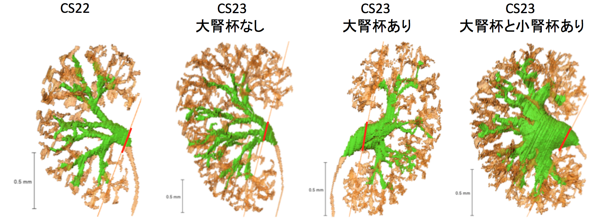
- 腎盂拡張はCS23内でみられる
- 分岐次数は極部で高い
34. Ishiyama H, Ishikawa A, Kitazawa H, Fujii S, Matsubayashi J, Yamada S, Takakuwa T, Branching morphogenesis of the urinary collecting system in the human embryonic metanephros, PLoS ONE 13(9): e0203623. https://doi.org/10.1371/journal.pone.0203623
Abstract
An elaborate system of ducts collects urine from all nephrons, and this structure is known as the urinary collecting system (UCS). This study focused on how the UCS is formed during human embryogenesis. Fifty human embryos between the Carnegie stage (CS) 14 and CS23 were selected from the Kyoto Collection at the Congenital Anomaly Research Center of Kyoto University, Japan. Metanephroses, including the UCS, were segmented on serial digital virtual histological sections. Three-dimensional images were computationally reconstructed for morphological and quantitative analyses. A CS timeline was plotted. It consisted of the 3-D structural morphogenesis of UCS and quantification of the total amount of end-branching, average and maximum numbers of generations, deviation in the metanephros, differentiation of the urothelial epithelium in the renal pelvis, and timing of the rapid expansion of the renal pelvis. The first UCS branching generation occurred by CS16. The average branching generation reached a maximum of 8.74 ± 1.60 and was already the twelfth in CS23. The total end-branching number squared between the start and the end of the embryonic period. UCS would reach the fifteenth branching generation soon after CS23. The number of nephrons per UCS end-branch was low (0.21 ± 0.14 at CS19, 1.34 ± 0.49 at CS23), indicating that the bifid branching occurred rapidly and that the formation of nephrons followed after. The renal pelvis expanded mainly in CS23, which was earlier than that reported in a previous study. The number of nephrons connected to the UCS in the expanded group (246.0 ± 13.2) was significantly larger than that of the pre-expanded group (130.8 ± 80.1) (P < 0.05). The urothelial epithelium differentiated from the zeroth to the third generations at CS23. Differentiation may have continued up until the tenth generation to allow for renal pelvis expansion. The branching speed was not uniform. There were significantly more branching generations in the polar- than in the interpolar regions (P < 0.05). Branching speed reflects the growth orientation required to form the metanephros. Further study will be necessary to understand the renal pelvis expansion mechanism in CS23. Our CS-based timeline enabled us to map UCS formation and predict functional renal capacity after differentiation and growth.