アクセスカウンター

2024年5月
 12345
6789101112
13141516171819
20212223242526
2728293031  

ヒト胎児の骨盤の性差は一次骨化が開始する受精後9週にはすでに存在する

概要

骨盤入口部、恥骨下角、骨盤出口部に性差あり

ヒトの骨盤には思春期以降、明確な性差が認められます。しかし、出生前の胎児期における骨盤の性差については、見解が一致していません。ヒトの骨盤は、軟骨原基が骨組織に置き換えられる軟骨内骨化によって形成されます。これまでの胎児期の性差の検討は、軟骨組織から骨組織への置き換え(一次骨化)がある程度進んだ、受精後20週以降が主に対象とされ、一次骨化が開始する受精後9週から対象とした解析はありませんでした。われわれは、京都大学医学研究科附属先天異常標本解析センター及び島根大学医学部解剖学講座が所蔵する、受精後9週から23週に相当する頭殿長 50~225mmのヒト胎児標本72体のMRI画像を取得し、様々な部位の計測と重回帰分析を行なって性差を検討しました。その結果、骨盤上口の前後径、恥骨下角、および坐骨棘間径と大骨盤横径の比に性差が認められました。従来考えられてきた時期よりも早く、一次骨化の開始期には既に骨盤に性差が存在することを示唆する本研究結果は、ヒト胎児骨盤の形態が男女で異なることを理解する上で重要な知見を提供するものです。

1.背景

思春期以降のヒトの骨盤に性差が見られることは広く知られています。では、具体的にいつからその性差が現れるのでしょうか。この点について、多くの研究者が興味を持ち、解析に取り組んできました。しかしながら、これまでの研究成果をみますと、ヒト胎児骨盤の性差の有無については意見が分かれています。さらに、これまでの研究は、主に受精後20週以降の胎児を対象としており、20週以前を対象とした解析は十分ではありませんでした。そのため、私たちは、外性器の肉眼観察から胎児の性別を判定できる最も早い時期であり、一次骨化が開始する受精後9週(頭殿長[注1] 50mm )からのヒト胎児標本を対象として、骨盤の性差を検討することにしました。

2.研究手法・成果

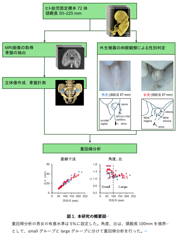
本研究はヒト胎児標本のMRI画像を取得した後、画像解析ソフトを用いて骨盤の立体像を作成しました(図1)。ヒト胎児標本は、京都大学医学研究科附属先天異常標本解析センターと島根大学医学部解剖学講座が保有する標本群を使用しました。この標本群は、世界最大規模の研究リソースとして知られており、その利用については京都大学大学院医学研究科・医学部及び医学部附属病院医の倫理委員会の承認のもとで研究が行われています。

ヒト胎児の性別は外生殖器の肉眼観察から判定しました。肉眼観察を用いた正確な性別判定は、頭殿長50mmの大きさの胎児から十分に可能です。本研究では、頭殿長が50mmから225mmまでのヒト胎児標本72体(男児;34体、女児;38体)のMRI画像を、前臨床用7T及び臨床用7T、3TMRI装置を用いて取得しました。作成した骨盤の立体像から、直線寸法や角度を21か所測定しました。また、直線寸法のデータを用いて計20個の比を算出しました。これらの計測データと重回帰分析を用いて性差を検討しました。

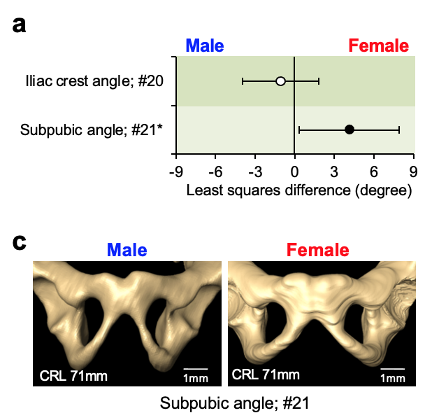
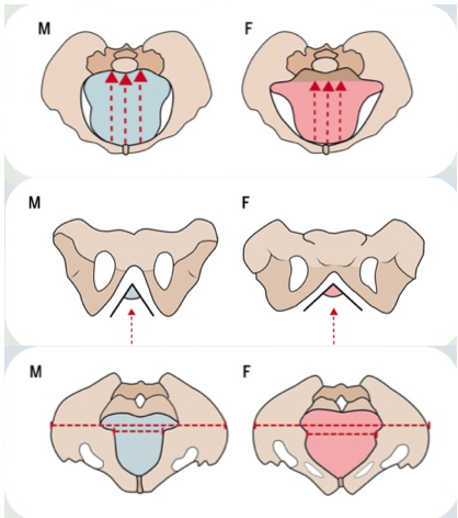
その結果、骨盤上口の前後径、前後径と横径の比、恥骨下角、坐骨棘間距離に対する大骨盤の横径(腸骨稜間距離、上前腸骨棘間距離)の比に有意な性差が確認されました(図2)。本研究成果から、これまでの報告よりも早い一次骨化が開始する受精後9週から、既にヒト胎児の骨盤には性差が存在することが示唆されます。

3.波及効果、今後の予定

本研究成果は、ヒト骨盤の性差に関する理解に大きく貢献し、さらに、骨盤形成の男女の違いについて、発生学や人類学などの様々な領域に新たな視点をもたらすことが期待されます。今回の検討では、なぜ一次骨化開始期から既に性差が存在するのかについては明らかにできていません。今後、ヒト骨盤の形態形成をより深く理解するために、一次骨化開始期以前の骨盤の性差の検討も含めて、より詳細に解析する必要があります。

<研究者のコメント>

本研究では、一次骨化が開始する受精後9週以降の希少なヒト胎児標本と、正確に立体情報を把握できる高解像度MRI画像を取得できたことで、従来では検討できなかった胎児期初期のヒト骨盤を検討し、性差の存在を証明することができました。本成果で得られた新しい知見は、「胎児期のヒトの骨盤では、性差は十分には分からない」と考えられてきた、これまでの定説を覆すという点で大きな価値を持つと考えます。(金橋 徹)

Kanahashi T, Matsubayashi J, Imai H, Yamada S, Otani H, Takakuwa T. Sexual dimorphism of the human fetal pelvis exists at the onset of primary ossification, Communications Biology, 2024, 7:538, https://doi.org/10.1038/s42003-024-06156-y

金橋先生の論文がCommun. Biol.に掲載

恥骨下角には性差がみられる

金橋先生の論文がCommunications Biologyに受諾されました。

ヒト胎児の骨盤形態の性差はこれまでの報告よりもずっと早く、一次骨化の開始時にすでに明らかであることを、統計学的に示しました。

*内容は、京大HP等で紹介されました

Kanahashi T, Matsubayashi J, Imai H, Yamada S, Otani H, Takakuwa T. Sexual dimorphism of the human fetal pelvis exists at the onset of primary ossification, Communications Biology, 2024, 7:538, https://doi.org/10.1038/s42003-024-06156-y

Abstract
Human adolescent and adult skeletons exhibit sexual dimorphism in the pelvis. However, the degree of sexual dimorphism of the human pelvis during prenatal development remains unclear. Here, we performed high-resolution magnetic resonance imaging-assisted pelvimetry on 72 human fetuses (males [M]: females [F], 34:38; 21 sites) with crown-rump lengths (CRL) of 50–225 mm (the onset of primary ossification). We used multiple regression analysis to examine sexual dimorphism with CRL as a covariate. Females exhibit significantly smaller pelvic inlet anteroposterior diameters (least squares mean, [F] 8.4 mm vs. [M] 8.8 mm, P = 0.036), larger subpubic angle ([F] 68.1° vs. [M] 64.0°, P = 0.034), and larger distance between the ischial spines relative to the transverse diameters of the greater pelvis than males. Furthermore, the sacral measurements indicate significant sex-CRL interactions. Our study suggests that sexual dimorphism of the human fetal pelvis is already apparent at the onset of primary ossification.

岩佐さんの修士論文がJ Anatomyに受諾

岩佐さんの腹直筋腱画についての論文がJ Anatomyに受諾されました。MRI_DTIを用いて腹直筋腱画形成過程を検討しました。

  • 胎児期早期の腹直筋腱画形成過程について、数、大きさ、タイプ、側方性、性別による検討を拡散テンソル画像を用いて行った。
  • 両側の腱性交差の平均数は3.1(範囲:2.0-4.0)であり、21%の検体では腱性交差は2つだけであった。
  • 腱性交差の形成における帯状の違いは、これまでの成人の研究で観察されたものとほぼ一致していた。拡散テンソル画像は、腱性交差部形成の早期分化を検出することができ有用であった。

Iwasa Y, KanahashiT, ImaiH, OtaniH, YamadaS, Takakuwa T. Formation of tendinous intersections in the human fetal rectus abdominis, J Anatomy 2024, in press, DOI: 10.1111/joa.14064

Abstract

Previous studies have poorly described the initial development process of the tendinous intersections of the rectus abdominis muscle (RAM). The present study aimed to observe the formation of tendinous intersections in the RAM during the early fetal period using diffusion tensor imaging (DTI). Fifteen human fetal specimens (crown-rump length [CRL]: 39.5–93.7 mm) were selected. Three-dimensional measurements revealed that Zone-4 (i.e., the zone between the pubic symphysis and the caudal base of the umbilical ring in the RAM) had a smaller width and was thicker than Zone-1 and Zone-2 (i.e., the zones between the costal arch and the cranial base of the umbilical ring) and Zone-3 (i.e., the zone at the umbilical ring). Characteristics of tendinous intersections in the RAM during the early fetal period were assessed according to number, size, type, laterality, and sex. The mean number of tendinous intersections on both sides was 3.1 (range: 2.0–4.0), and 21% of specimens had only two tendinous intersections, which was higher than that reported in previous adult studies. The present data suggest that the formation of tendinous intersections was still in progress in specimens with two tendinous intersections in the RAM and that the third tendinous intersection was formed in Zone-2. Ordinal logistic regression via generalized estimating equations revealed that the odds for a higher type of tendinous intersections in Zone-1 and Zone-2 were significantly higher than those in Zone-4 (adjusted odds ratio: 14.85, 8.84). The odds for the presence of incomplete types (tendinous intersections that could not completely transverse the RAM) in Zone-3 were significantly higher than those in Zone-1 (adjusted odds ratio: 7.4). The odds for missing tendinous intersections in Zone-4 were significantly higher than those in Zone-1 (adjusted odds ratio: 20.5). These zonal differences in the formation of tendinous intersections were consistent with those observed in previous adult studies. In this study, DTI detected tendinous intersections in a sample with a CRL of 45.8 mm (approximately 11 weeks of gestation), which is earlier than that in previous histological findings, indicating that the RAM does not have mature tendinous intersections until the 17th week of gestation. In conclusion, DTI could detect the premature differentiation of tendinous intersection formation. Our data may aid in elucidating the developmental processes of tendinous intersections in the RAM.

5/18;13時から大学院説明会を開催

総合医療科学コース 大学院説明会を5/18;13-15時 on line開催します。
・参加希望者は5月15日(水)までに以下のサイトで事前登録をお願いします.

腎臓の高さの左右差についてがCongenital Anomaliesに掲載

腎臓の高さの左右差についてがCongenital Anomaliesに掲載されました。

胚子期の腎臓の上昇、回転についてはこれまでよく知られていましたが、腎臓の高さの左右差については不明でした。本研究では、胚子期に左右腎臓の高さに違いはなく、ともに上昇すること、胎児期初期になると右側が左側よりも高くなることを示しました。胎児期の腎臓上昇の様子は、胚子期とは傾向が異なり別の機序を考える必要があります。

Ishiyama-Takara H, Matsubayashi J, Yamada S, Tetsuya Takakuwa T, Height difference between the right and left metanephroi during early human fetal development, Congenit Anom 64(3) 164-166, 2024.