


金橋先生の論文がCommunications Biologyに受諾されました。
ヒト胎児の骨盤形態の性差はこれまでの報告よりもずっと早く、一次骨化の開始時にすでに明らかであることを、統計学的に示しました。
*内容は、京大HP等で紹介されました
Kanahashi T, Matsubayashi J, Imai H, Yamada S, Otani H, Takakuwa T. Sexual dimorphism of the human fetal pelvis exists at the onset of primary ossification, Communications Biology, 2024, 7:538, https://doi.org/10.1038/s42003-024-06156-y
Abstract
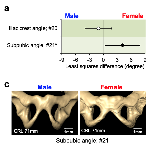
Human adolescent and adult skeletons exhibit sexual dimorphism in the pelvis. However, the degree of sexual dimorphism of the human pelvis during prenatal development remains unclear. Here, we performed high-resolution magnetic resonance imaging-assisted pelvimetry on 72 human fetuses (males [M]: females [F], 34:38; 21 sites) with crown-rump lengths (CRL) of 50–225 mm (the onset of primary ossification). We used multiple regression analysis to examine sexual dimorphism with CRL as a covariate. Females exhibit significantly smaller pelvic inlet anteroposterior diameters (least squares mean, [F] 8.4 mm vs. [M] 8.8 mm, P = 0.036), larger subpubic angle ([F] 68.1° vs. [M] 64.0°, P = 0.034), and larger distance between the ischial spines relative to the transverse diameters of the greater pelvis than males. Furthermore, the sacral measurements indicate significant sex-CRL interactions. Our study suggests that sexual dimorphism of the human fetal pelvis is already apparent at the onset of primary ossification.