アクセスカウンター

2024年12月
 1
2345678
9101112131415
16171819202122
23242526272829
3031  

第23回日本心臓血管発生研究会で発表

第23回日本心臓血管発生研究会しました(2024.11.13-14淡路島)

藤井 瀬菜, 福井 成美, 金橋 徹, 松林 潤, 今井 宏彦, 米山 明男, 大谷 浩, 山田 重人, 高桑 徹也 
ヒト胚子期から胎児初期における肺静脈と左心耳の形態形成 Morphogenesis of the pulmonary vein and left atrial appendage in human embryos and early fetuses
福井さんの修士研究を紹介いたしました。

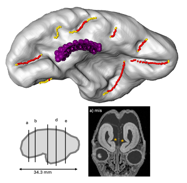
ヒトの左心房および肺静脈は、原始心房由来の心房前壁、左心耳、心房中隔と、大部 分が肺静脈に由来する後壁、肺静脈で構成されている。受精後4週ころ、左房後壁の一 部が突出し、肺静脈の原基である共通肺静脈を形成する。共通肺静脈は両側に分岐を 形成し、4本の肺静脈を形成したのち、左心房壁に順次取り込まれ融合する。この融合 の時期に関する記載は、受精後8~11週と様々であり統一されていない。そこで本研究 は、ヒトの胚子期から胎児期における、左心耳を含む左心房と肺静脈を形態学的およ び定量的に評価した。対象は、Carnegie stage (CS)16~23(受精後約6~8週に相 当)のヒト胚子標本23体とヒト胎児期初期の標本19体の、phase-contrast X-ray computed tomographyおよびmagnetic resonance imaging画像とした。画像解析 ソフトウェアAmiraを用いて、左心房と肺静脈を立体再構成した。形態学的評価におい て、各個体にて心膜が反転する位置を心膜腔の境界として定義し、共通肺静脈や4本の 肺静脈(左上、左下、右上、右下肺静脈)と心膜との位置関係を同定した。その結 果、CS19以降の4個体を除く全個体において、4本すべての肺静脈の周縁で心膜が反転 していた。この結果は、心臓のdorsal mesocardiumの接続が退縮したときに、2つの 肺静脈(右肺静脈と左肺静脈)と4つの肺静脈で心膜が反転する位置がほぼ同時に決定 することを示す。また、3次元立体像より、4本の肺静脈が左心房へ向かって外側から 内側へ接線方向に接続する様子を観察した。右肺静脈の左心房への侵入角度は、左肺 静脈よりも大きかった。定量的評価として、(1)各肺静脈間の距離、(2)静脈内腔の断 面積と扁平率、(3)左心耳の開口部の断面積と扁平率、(4)左心耳と左心房の各容量お よび容量比を算出した。(1)上肺静脈と下肺静脈の間の距離は、左肺静脈と右肺静脈の 間の距離よりも短かった。(2)4本の肺静脈のうち、左上肺静脈は断面積が最も小さ く、最も扁平化した形状であった。他の3本は断面積と扁平率に差を認めず、類似の形 態であった。(3)左心耳の開口部は、発生に伴い断面積が大きくなると同時に平坦にな る傾向を認めた。(4)左心耳は中心部の容積が大きく、発生が進むにつれて葉状構造と して左室を覆うように拡大していた。左心房全体の容積^(1/3)は、頭殿長にほぼ比例 して増加した。本研究は、ヒトの胚子期後期から胎児期初期にかけて、肺静脈の左心 房壁への取り込みや左心耳の形態形成を定性的、定量的に明らかにした。また、これ まで注目されていなかった肺静脈の心膜腔への取り込み時期についても明らかにし た。

熊谷さんの卒業研究がAnat Recに受諾

熊谷さんの卒業研究がAnat Recに受諾されました。1次脳溝(脳のしわ)が、どのように最初できてくるかを検討しました。

  • 妊娠11週から16週までの33のサンプルから得られた高解像度の磁気共鳴画像(頭殿長<130 mm)を使用して、脳溝形成の出現時期と形態学的および形態計測学的特徴を特定。
  • 検出時期は、脳梁では12週未満、海馬、鳥骨、頭頂後頭溝では13週未満、円形島状溝では15週未満、嗅溝はときおり検出され、帯状溝は特定されていない。円形の島状溝が徐々に現れ形を変える
  • 測定された溝はどれも頭殿長と相関しない。溝はある程度の距離にわたって同時に発生する可能性あり。 出生前診断や神経発達障害の研究に大きな影響を与える可能性

70. Kumagai M, Kanahashi M,  Matsubayashi J, Imai H, Otani H, Takakuwa T.
Primary sulci formation in human cerebral cortex development. Anat Rec (Hoboken) 2025, in press 

Abstract

We aimed to determine the timing of appearance and the morphologic and morphometric features of the initial human cerebral sulcal formation. Using high-resolution magnetic resonance images obtained from 33 samples between 11 and 16 weeks (w) of gestation (crown-rump length <130 mm), the cerebral surface and internal structures on serial two-dimensional planes and all possible sulci on three-dimensional reconstructions were marked, allowing comparison of the positions of the sulci in the samples and inter-samples. Our method provided accurate conclusions regarding the timing of sulcal formation. Detection timing was as early as and earlier than those in previous studies using anatomical dissection and magnetic resonance imaging (MRI), respectively: <12 w for the callosum, <13 w for the hippocampal, calcarine, and parieto-occipital sulci, and <15 w for the lateral sulcus. Occasionally, an olfactory sulcus was detected. However, the cingulate sulcus could not be definitely identified. The lateral sulcus gradually appeared and changed shape. The lengths of the left and right sides of the olfactory sulci and the left side of the hippocampal sulcus increased linearly with the CRL. The length of the right side of the hippocampal sulcus and the left and right sides of the calcarine, parieto-occipital, and not determined_a sulci did not increase with the CRL The depth of the all sulci, except for the parieto-occipital sulci, increased linearly with the CRL. The sulci might not arise as if they elongate gradually but arise simultaneously over some distance. We determined the timing of the initial sulcal formation using high-resolution MRI. Our findings may significantly impact prenatal diagnosis and research on neurodevelopmental disorders.