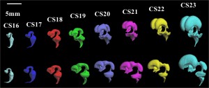
中島さんの修士論文がCongenit Anomに掲載されました。

- 胚子期の脳神経管と脳室の平面・立体像を作成し検討。
- 脳胞背側長、腹側長はCS18からCS23の間は増加し、背側の方が増加量が4.2倍高い
- 前脳の増加量が最も高く背側は腹側の3倍以上
- これらの結果は、胚子期の脳胞発生の特徴である終脳の急速な増大を反映
2.Nakashima T, Hirose A, Yamada S, Uwabe C, Kose K, Takakuwa T, Morphometric analysis of the brain vesicles during the human embryonic period by magnetic resonance microscopic imaging, Congenit Anom (Kyoto). 2012 Mar;52(1):55-8, doi; 10.1111/j.1741-4520.2011.00345.x
ABSTRACT
The development of the brain vesicles between Carnegie stages (CS) 17 and 23 was analyzed morphometrically using 177 magnetic resonance image data derived from the Kyoto Collection of Human Embryos. Whole embryonic volume was 106.55 ± 21.08 mm3 at CS17, exponentially increasing to CS23 when it reached 1357.28 ± 392.20 mm3. Length of brain vesicles was 29.83 ± 2.52 mm at CS17, increased almost linearly and reached 49.31 ± 6.66 mm at CS23. The rate of increase was approximately 4.2 times higher on the dorsal side than on the ventral side. The increase in the length of the brain vesicles resulted mainly from that of the prosencephalon, and the rate of increase was three times higher on the dorsal side than on the ventral side of the prosencephalon.


