


")
白石くんの修士論文”Morphogenesis of lateral choroid plexus during human embryonic period” -ヒト胚子期における側脳室脈絡叢の形態、組織学的研究-がAnatomical Recordに掲載されました。
- CS18 – CS23 における脈絡叢 (CP) の形成を検討。
- CS19 ;CP の原基、尾側に成長した小さな三日月形の塊として検出
- CS20; 多数の起伏のある表面
- CS21 で不規則な膨らみを形成、全方向に成長
- CS23 で尾側表面に 2 つの深裂、 3 つの大きなクラスターを形成
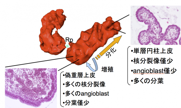
- 近位領域は未分化な上皮、血管芽細胞が増殖、遠位領域は分化、分葉化した組織を観察
6. Shiraishi N, Nakashima T, Yamada S, Uwabe C, Kose K, Takakuwa T, Morphogenesis of lateral choroid plexus during human embryonic period, Anat Rec (Hoboken). 2013 Apr;296(4):692-700. doi: 10.1002/ar.22662
Abstract
The morphological and histological changes of the choroid plexus (CP) during Carnegie stage (CS) 18 and CS23 were presented, based on magnetic resonance imaging data and histological serial section of human embryos from the Kyoto Collection of Human Embryos. The primordium of the CP was initially detected as a small lump at CS19 that grew caudally, so that the CP became crescent shaped. It developed in all directions after CS21, as the dorsal and frontal growth also became prominent. The CP formed a number of undulating surfaces at CS20, irregular bulges at CS21, and then three large clusters with two deep fissures on the caudal surface at CS23. The mean volume of the CP was 0.282±0.141 mm3 at CS19; it reached 16.8±8.77 mm3 at CS23. Additionally, the histology was different depending on the regions of the CP at all stages after CS20. The epithelium and angioblasts in the center of the stroma were proliferated in the proximal region, whereas the epithelium was differentiated and lobulated in the distal region where the blood vascular system was organized. The histological differentiation was mapped on the CP reconstructed from histological serial sections. The data suggested the correlation between morphological information obtained from magnetic resonance data sets and distribution of the differentiation. With the help of morphological analysis and histological findings, we have been able to categorize each CP into specific stages. These findings will be useful in clinical evaluation of development during the embryonic period.