


吉田さんの卒論がCongenit Anomに掲載されました。胎児の脳溝形成の様子をGyrification Indexを用いて解析しました。また、同号の表紙に、論文のFigureが採用されました。
- 16 -40 週の大脳の脳溝形成について、GI値を用いて検討
- GI 値は、体重500g(CRL 200 mm )まで約 1.0(脳溝なし)
- その後、GI値は体重と CRL に相関して増加(脳溝形成あり)
- 妊娠21週で一次脳溝とGIのピークが対応
- 31-40週数で前頭側頭領域よりも頭頂後頭領域でGI値は増加
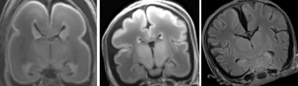
Gyrification index (GI) of brain slices in a left lateral view of the brain in a 1409g fetus. The line graph above the brain shows the GI of each slice below it. The GI of the whole hemisphere changed in the rostro-caudal dimension. Several maxima of the GI curve appeared in relation to gross landmarks. Further details can be seen in the article by Yoshida et al. in this issue.
27. Yoshida R, Koichi Ishizu K, Yamada S, Chigako Uwabe C, Okada T, Togashi K, Takakuwa T, The dynamics of gyrification in the human cerebral cortex during development, Congenit Anom, 57 (1) 8-14, 2017, DOI: 10.1111/cga.12179, 10.1111/cga.12181
Abstract
This study quantitatively characterized cortical gyrus folding over human neocortical development by calculating the gyrification index (GI) in 22 human fetal specimens from 16 to 40 weeks with magnetic resonance imaging data. GI values remained constant at approximately 1.0 until the fetal specimens reached 500 g body weight and 200 mm crown-rump length (CRL), respectively, and then increased in correlation with the body weight and CRL. The rostrocaudal GI distribution in the cerebral cortex revealed a correspondence of GI peaks with indentations of early-generated primary sulci at 21 weeks of gestation and more frequently increased GI values in the parieto-occipital region than in the fronto-temporal region at 31 and 40 weeks of gestation. These results provide a quantitative reference set for gyrification in normal human cortical development, which may help reveal the mechanism of neurodevelopmental disorders.