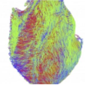
西谷さんの修士論文、鳥居さんの卒業論文;胎児心臓のDT-MRIによる解析がJAHAに掲載されました。
- 従来の報告より8週以上若い時期のヒトの心筋走行を高解像度DT-MRIデータを用いて検出しました。
- ヒトの心筋の走行の配置、螺旋状の角度等は、成人とすでに同様であることから、心筋構築の青写真は胚子期末(CS20-23, GSA8W)には、すでに形成されていることをから明らかにしました。
- データは古典的な心筋バンド説(心筋はひとつながりのバンド状のものが立体的に折り畳まれて形成されている)を否定しました。
本研究成果は、京都大学HP(トピックス)でも紹介されました。
ヒト胎児心臓の心筋線維方向を追跡 -受精後8週の心筋線維は成人と同じ配列をする- 外部リンク[京都大学HP(トピックス)]
2020/10/13 医療NEWSにて,ヒト胎児の心臓、受精後8週で心筋線維の配列が成人と同様であることが判明-京大ほか が紹介されました.
46. Nishitani S, Torii N, Imai H, Haraguchi R, Yamada S, Takakuwa T, Development of helical myofiber tracts in the human fetal heart: Analysis of myocardial fiber formation in the left ventricle from the late human embryonic period using diffusion tensor magnetic resonance imaging. Journal of the American Heart Association, 2020, 19(9) doi:10.1161/JAHA.120.016422
Abstract
Background
Detection of the fiber orientation pattern of the myocardium using diffusion tensor magnetic resonance imaging lags ≈12 weeks of gestational age (WGA) behind fetal myocardial remodeling with invasion by the developing coronary vasculature (8 WGA). We aimed to use diffusion tensor magnetic resonance imaging tractography to characterize the evolution of fiber architecture in the developing human heart from the later embryonic period.
Methods and Results
Twenty human specimens (8–24 WGA) from the Kyoto Collection of Human Embryos and Fetuses, including specimens from the embryonic period (Carnegie stages 20–23), were used. Diffusion tensor magnetic resonance imaging data were acquired with a 7T magnetic resonance system. Fractional anisotropy and helix angle were calculated using standard definitions. In all samples, the fibers ran helically in an organized pattern in both the left and right ventricles. A smooth transmural change in helix angle values (from positive to negative) was detected in all 16 directions of the ventricles. This feature was observed in almost all small (Carnegie stage 23) and large samples. A higher fractional anisotropy value was detected at the outer side of the anterior wall and septum at Carnegie stage 20 to 22, which spread around the ventricular wall at Carnegie stage 23 and in the early fetal samples (11–12 WGA). The fractional anisotropy value of the left ventricular walls decreased in samples with ≥13 WGA, which remained low (≈0.09) in larger samples.
Conclusions
From the human late embryonic period (from 8 WGA), the helix angle arrangement of the myocardium is comparable to that of the adult, indicating that the myocardial structure blueprint, organization, and integrity are already formed.


