


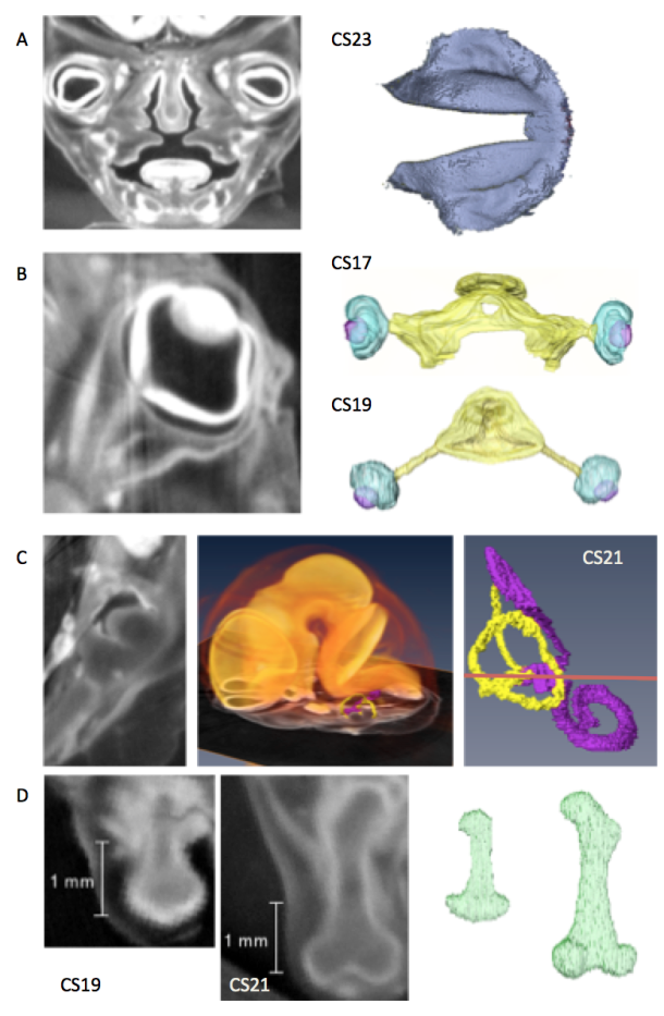
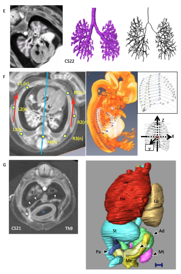
Figure legends
Representative studies using CT images acquired from the Kyoto collection.
CT planes and reconstructed images were provided.
A) Coronal plane of the face and reconstruction of the secondary palate just before fusion (CS23)[1].
B) Sagittal plane of the eyeball and the reconstructions of the eyeball with the optic nerve (CS17 and CS19)[2].
C) Transverse plane of the temporal bone and reconstruction of the inner ear with enlarged brain and inner ear (CS21)[3]
D) Sagittal plane of the thigh and reconstructions of the femur (CS19, CS21)[4]
E) transverse plane of the thorax and reconstructions and median line of the bronchial tree (CS22)[5].
F) transverse plane of the thorax and reconstruction of the whole embryo, thoracic cage, and analysis of the reconstructed image (CS22)[6].
G) Transverse plane of the thorax and abdomen and reconstructions of the thoracic and abdominal organs (CS21)[7]. In this sample, the liver is defective, and the position of organs such as the stomach, pancreas, and heart has been changed.
1. Nohara A, Owaki N, Matsubayashi J, Katsube M, Imai H, Yoneyama A, Yamada S, Kanahashi T, Takakuwa T. Morphometric analysis of secondary palate development in human embryos. J Anat, 2022, 241(6), 1287-1302, 2022, DOI:10.1111/joa.13745
2. Osaka M, Ishikawa A, Yamada S, Uwabe C, Imai H, Matsuda T, Yoneyama A, Takeda T, Takakuwa T, Positional changes of the ocular organs during craniofacial development, Anat Rec (Hoboken) 300(12), 2107–2114, 2017 DOI: 10.1002/ar.23588
3. Toyoda S, Shiraki N, Yamada S, Uwabe C, Imai H, Matsuda T, Yoneyama A, Takeda T, Takakuwa T, Morphogenesis of the inner ear at different stages of normal human development. Anat Rec (Hoboken) 298:2081–2090 (2015), doi: 10.1002/ar.23268
4. Suzuki Y, Matsubayashi J, Ji X, Yamada S, Yoneyama A, Imai H, Matsuda T, Aoyama T, Takakuwa T Morphogenesis of the femur at different stages of normal human development, PLoS ONE, 14(8): e0221569. https://doi.org/10.1371/journal. pone.0221569
5. Fujii S, Muranaka T, Matsubayash J, Yamada S, Yoneyama A, Takakuwa T. The bronchial tree of the human embryo: an analysis of variations in the bronchial segments. J Anat 2020, 237, 311-322. doi: 10.1111/joa.13199.
6. Okuno K, Ishizu K, Matsubayashi J, Fujii S, Sakamoto R, Ishikawa A, Yamada S, Yoneyama A, Takakuwa T. Rib cage morphogenesis in the human embryo: A detailed three-dimensional analysis. Anat Rec (Hoboken) 2019, 302, 2211-2223, doi: 10.1002/ar.24226
7. Kanahashi T, Yamada S, Tanaka M, Hirose A, Uwabe C, Kose K, Yoneyama A, Takeda T, Takakuwa T, A novel strategy to reveal the latent abnormalities in human embryonic stages from a large embryo collection, Anat Rec (Hoboken) 299,8-24,2016