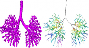
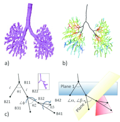
44. Fujii S, Muranaka T, Matsubayash J, Yamada S, Yoneyama A, Takakuwa T. The bronchial tree of the human embryo: an analysis of variations in the bronchial segments. J Anat 2020, 237, 311-322. doi: 10.1111/joa.13199.
48. Fujii S, Muranaka T, Matsubayashi J, Yamada S, Yoneyama A, Takakuwa T. Bronchial tree of the human embryo: categorization of the branching mode as monopodial and dipodial, PLoS One 16; e0245558, 2021, https://doi.org/10.1371/journal.pone.0245558
Gyrification index (GI) of brain slices in a left lateral view of the brain in a 1409g fetus. The line graph above the brain shows the GI of each slice below it. The GI of the whole hemisphere changed in the rostro-caudal dimension. Several maxima of the GI curve appeared in relation to gross landmarks. Further details can be seen in the article by Yoshida et al. in this issue.
27. Yoshida R, Koichi Ishizu K, Yamada S, Chigako Uwabe C, Okada T, Togashi K, Takakuwa T, The dynamics of gyrification in the human cerebral cortex during development, Congenit Anom, 57 (1) 8-14, 2017, DOI: 10.1111/cga.12179, 10.1111/cga.12181
Abstract
This study quantitatively characterized cortical gyrus folding over human neocortical development by calculating the gyrification index (GI) in 22 human fetal specimens from 16 to 40 weeks with magnetic resonance imaging data. GI values remained constant at approximately 1.0 until the fetal specimens reached 500 g body weight and 200 mm crown-rump length (CRL), respectively, and then increased in correlation with the body weight and CRL. The rostrocaudal GI distribution in the cerebral cortex revealed a correspondence of GI peaks with indentations of early-generated primary sulci at 21 weeks of gestation and more frequently increased GI values in the parieto-occipital region than in the fronto-temporal region at 31 and 40 weeks of gestation. These results provide a quantitative reference set for gyrification in normal human cortical development, which may help reveal the mechanism of neurodevelopmental disorders.
30. Takakuwa T, 3D analysis of human embryos and fetuses using digitized datasets from the Kyoto Collection, Anat Rec 2018, 301,960-969 doi: 10.1002/ar.23784 (英文で読む)
ABSTRACT
Three-dimensional (3D) analysis of the human embryonic and early-fetal period has been performed using digitized datasets obtained from the Kyoto Collection, in which the digital datasets play a primary role in research. Datasets include magnetic resonance imaging (MRI) acquired with 1.5 T, 2.35 T, and 7 T magnet systems, phase-contrast X-ray computed tomography (CT), and digitized histological serial sections. Large, high-resolution datasets covering a broad range of developmental periods obtained with various methods of acquisition are key elements for the studies. The digital data have gross merits that enabled us to develop various analysis. Digital data analysis accelerated the speed of morphological observations using precise and improved methods by providing a suitable plane for a morphometric analysis from staged human embryos. Morphometric data are useful for quantitatively evaluating and demonstrating the features of development and for screening abnormal samples, which may be suggestive in the pathogenesis of congenital malformations. Morphometric data are also valuable for comparing sonographic data in a process known as “sonoembryology.” The 3D coordinates of anatomical landmarks may be useful tools for analyzing the positional change of interesting landmarks and their relationships during development. Several dynamic events could be explained by differential growth using 3D coordinates. Moreover, 3D coordinates can be utilized in mathematical analysis as well as statistical analysis. The 3D analysis in our study may serve to provide accurate morphologic data, including the dynamics of embryonic structures related to developmental stages, which is required for insights into the dynamic and complex processes occurring during organogenesis. Anat Rec, 301:960–969, 2018.
26. Ozeki-Satoh M, Ishikawa A, Yamada S, Uwabe C, Takakuwa T. Morphogenesis of the Middle Ear Ossicles and Spatial Relationships with the External and Inner Ears during the Embryonic Period, Anat Rec 299:1325–1337, 2016, DOI 10.1002/ar.23457
Abstract
We describe the three-dimensional morphogenesis of the middle ear ossicles (MEOs) according to Carnegie stage (CS) in human embryos. Seventeen samples including 33 MEOs from CS18 to 23 were selected from the Kyoto Collection. The primordia of the MEOs and related structures were histologically observed and three-dimensionally reconstructed from digital images. The timing of chondrogenesis was variable among structures. The stapes was recognizable as a vague condensation of the mesenchymal cells in all samples from CS18, whereas the malleus and incus were recognizable at CS19. Chondrogenesis of all MEOs was evident in all samples after CS21. The chondrocranium was recognizable in all samples by CS18, and the perichondrium border of the auricular cartilage and otic capsule was distinct in all samples at CS23. At CS19, the MEOs were positioned in the anterior to posterior direction, following the order malleus, incus, stapes, which adjusted gradually during development. The MEOs connected in all samples after CS22. The stapes was located close to the vestibular part of the inner ear, although the basal part was not differentiated into the “footplate” form, even at CS23. The handles of the malleus were close to the tubotympanic recess at CS23, but were distant from the external auditory meatus. Determining the timeline of the formation of MEOs and connection of the external and inner ears can be informative for understanding hearing loss caused by failure of this connection. These data may provide a useful standard for morphogenesis, and will contribute to distinguishing between normal and abnormal MEO development.
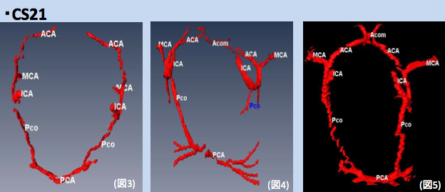
25. Takakuwa T, Koike T, Muranaka T, Yamada S, Uwabe C. 2016. Formation of the circle of Willis during human embryonic development. Congenit Anom (Kyoto) 2016; 56, 233–236, DOI: 10.1111/cga.12165
Abstract
The circle of Willis (CW) is a circulatory anastomosis that supplies blood to the brain and adjacent structures. We examined the timing of formation of CW in 20 Japanese human embryo samples by using 3-dimensional reconstruction of serial histological sections. The CW was closed in 1 (n = 6), 2 (n = 8), 2 (n = 3) and 2 (n = 3) samples at Carnegie stages 20, 21, 22, and 23, respectively. The CW was unclosed in 13 samples (unclosed at ACOM alone, 6 samples; ACOM and bilateral P1, 4; left PCOM and right P1, 1; right PCOM and right P1, 1; ACOM and left PCOM, 1). It was difficult to predict whether the circle would close during further development, as such variations frequently exist in adults.