26. Ozeki-Satoh M, Ishikawa A, Yamada S, Uwabe C, Takakuwa T. Morphogenesis of the Middle Ear Ossicles and Spatial Relationships with the External and Inner Ears during the Embryonic Period, Anat Rec 299:1325–1337, 2016, DOI 10.1002/ar.23457
Abstract
We describe the three-dimensional morphogenesis of the middle ear ossicles (MEOs) according to Carnegie stage (CS) in human embryos. Seventeen samples including 33 MEOs from CS18 to 23 were selected from the Kyoto Collection. The primordia of the MEOs and related structures were histologically observed and three-dimensionally reconstructed from digital images. The timing of chondrogenesis was variable among structures. The stapes was recognizable as a vague condensation of the mesenchymal cells in all samples from CS18, whereas the malleus and incus were recognizable at CS19. Chondrogenesis of all MEOs was evident in all samples after CS21. The chondrocranium was recognizable in all samples by CS18, and the perichondrium border of the auricular cartilage and otic capsule was distinct in all samples at CS23. At CS19, the MEOs were positioned in the anterior to posterior direction, following the order malleus, incus, stapes, which adjusted gradually during development. The MEOs connected in all samples after CS22. The stapes was located close to the vestibular part of the inner ear, although the basal part was not differentiated into the “footplate” form, even at CS23. The handles of the malleus were close to the tubotympanic recess at CS23, but were distant from the external auditory meatus. Determining the timeline of the formation of MEOs and connection of the external and inner ears can be informative for understanding hearing loss caused by failure of this connection. These data may provide a useful standard for morphogenesis, and will contribute to distinguishing between normal and abnormal MEO development.
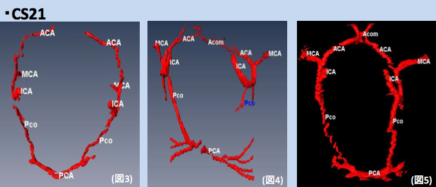
25. Takakuwa T, Koike T, Muranaka T, Yamada S, Uwabe C. 2016. Formation of the circle of Willis during human embryonic development. Congenit Anom (Kyoto) 2016; 56, 233–236, DOI: 10.1111/cga.12165
Abstract
The circle of Willis (CW) is a circulatory anastomosis that supplies blood to the brain and adjacent structures. We examined the timing of formation of CW in 20 Japanese human embryo samples by using 3-dimensional reconstruction of serial histological sections. The CW was closed in 1 (n = 6), 2 (n = 8), 2 (n = 3) and 2 (n = 3) samples at Carnegie stages 20, 21, 22, and 23, respectively. The CW was unclosed in 13 samples (unclosed at ACOM alone, 6 samples; ACOM and bilateral P1, 4; left PCOM and right P1, 1; right PCOM and right P1, 1; ACOM and left PCOM, 1). It was difficult to predict whether the circle would close during further development, as such variations frequently exist in adults.
23. Kobayashi A, Ishizu K, Yamada S, Uwabe C, Kose K, Takakuwa T, Morphometric human embryonic brain features according to developmental stage, Prenatal Diagnosis, 36:338–345, 2016, DOI: 10.1002/pd.4786. DOI: 10.1002/pd.4818
Abstract
Objectives
The present study investigated linear, area, and volume measurements of human brain samples according to Carnegie stages (CS) in an attempt to select suitable morphometric features that reflect embryonic development.
Methods
Using magnetic resonance imaging, we measured seven linear segments, three separate areas, and three regional volumes in 101 samples between CS13 and 23. Brain volume was determined via manual segmentation of the magnetic resonance image, whereby a formula was generated to estimate the volume of each linear measurement.
Results
All parameters correlated with crown-rump length. Bitemporal length and mesencephalic height increased linearly according to the CS, and a high correlation between bitemporal length and both whole-brain (r = 0.98) and prosencephalon (r = 0.99) volumes was found when brain cavity volume was excluded.
Conclusion
Morphometric data related to human embryonic stages are valuable for correcting and comparing sonographic data. The present approach may contribute to improvements in prenatal diagnostics by enabling the selection of more suitable measurements during early embryonic stages.
22. Ueno S, Yamada S, Uwabe C, Männer J, Shiraki N, Takakuwa T, The digestive tract and derived primordia differentiate by following a precise timeline in human embryos between Carnegie stages 11 and 13, Anatomical Rec 2016, 299(4), 439-449, DOI: 10.1002/ar.23314
ABSTRACT
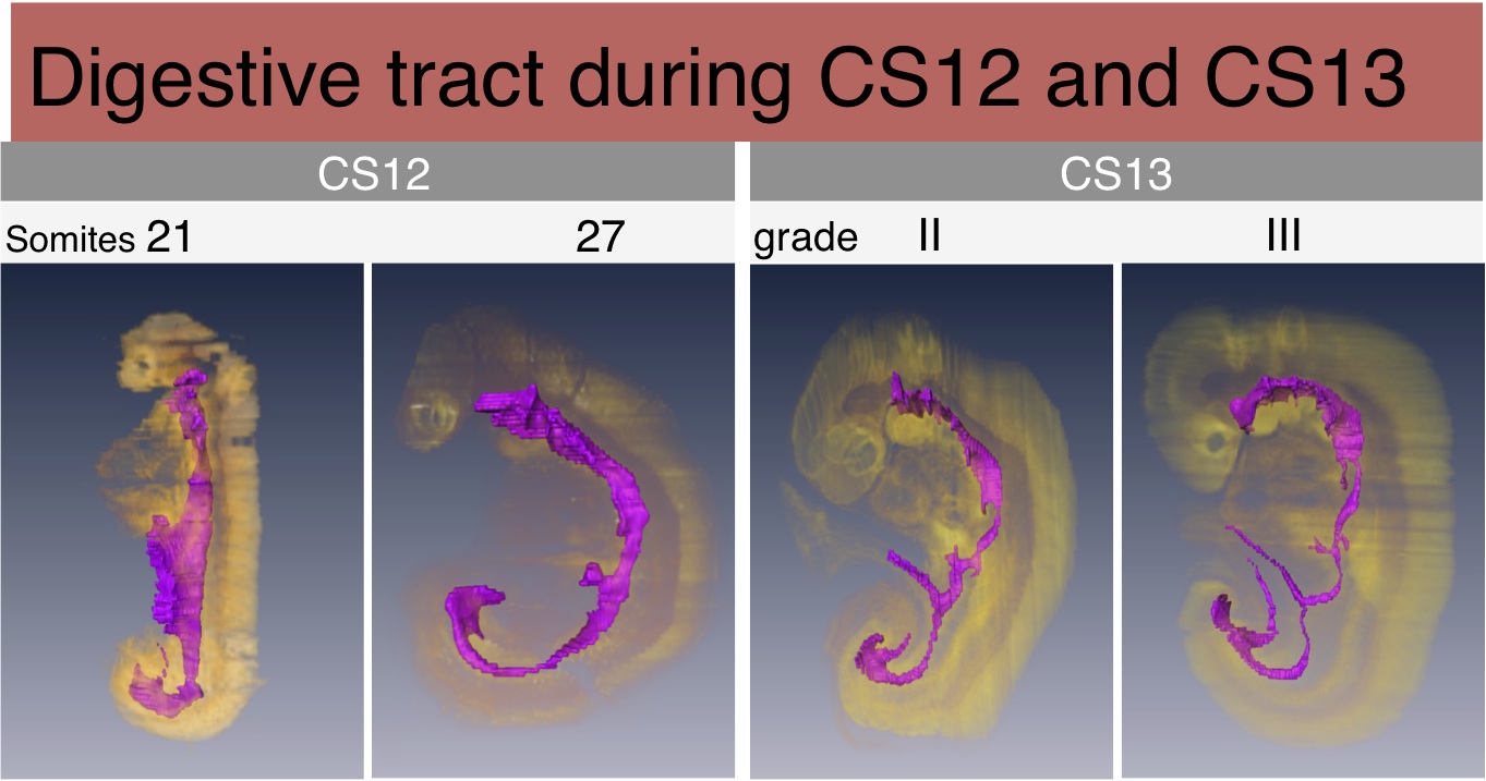
The precise mechanisms through which the digestive tract develops during the somite stage remain undefined. In this study, we examined the morphology and precise timeline of differentiation of digestive tract-derived primordia in human somite-stage embryos. We selected 37 human embryos at Carnegie Stage (CS) 11–CS13 (28–33 days after fertilization) and three-dimensionally analyzed the morphology and positioning of the digestive tract and derived primordia in all samples, using images reconstructed from histological serial sections. The digestive tract was initially formed by a narrowing of the yolk sac, and then several derived primordia such as the pharynx, lung, stomach, liver, and dorsal pancreas primordia differentiated during CS12 (21–29 somites) and CS13 (≥ 30 somites). The differentiation of four pairs of pharyngeal pouches was complete in all CS13 embryos. The respiratory primordium was recognized in ≥ 26-somite embryos and it flattened and then branched at CS13. The trachea formed and then elongated in ≥ 35-somite embryos. The stomach adopted a spindle shape in all ≥ 34-somite embryos, and the liver bud was recognized in ≥ 27-somite embryos. The dorsal pancreas appeared as definitive buddings in all but three CS13 embryos, and around these buddings, the small intestine bent in ≥ 33-somite embryos. In ≥ 35-somite embryos, the small intestine rotated around the cranial-caudal axis and had begun to form a primitive intestinal loop, which led to umbilical herniation. These data indicate that the digestive tract and derived primordia differentiate by following a precise timeline and exhibit limited individual variations.
21. Ozeki-Sato M, Yamada S, Uwabe C, Ishizu K, Takakuwa T, Correlation of external ear auricle formation with staging of human embryos, Congenit Anom (Kyoto) 56, 86-90, 2016, DOI: 10.1111/cga.12140, . (概要),
Abstract
The formation of auricles in human embryos was evaluated between Carnegie stage (CS)19 and CS23, and the findings were correlated across the stages. The auricle was categorized into 11 steps according to Streeter’s criteria with modifications. Mesenchyme cell condensation was observed at Step 7, and two layers of cartilage consisting of the auricle were recognized at Step11. The representative steps at each CS shifted from Step 3 to Step11 during CS16 and CS23, although several steps overlapped between adjacent CSs. These results indicate that observations of the auricle between CS19 and CS23 may be utilized for determining embryo staging as convincing supportive evidence of external features reflecting the internal histological structure, although other findings should also be taken into account.