Fujii S, Muranaka T, Matsubayashi J, Yamada S, Yoneyama A, Takakuwa T. Bronchial tree of the human embryo: Examination based on a mammalian model. J Anatomy 2024, 244, 159-169 http://doi.org/10.1111/joa.13946 .
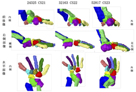
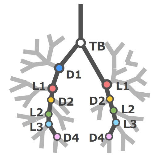
The symmetry of right and left bronchi, proposed in a previous comparative anatomical study as the basic model of the mammalian bronchial tree, was examined to determine if it applied to the embryonic human bronchial tree. Imaging data of 41 human embryo specimens at Carnegie stage (CS) 16–23 (equivalent to 6–8 weeks after fertilization) belonging to the Kyoto collection were obtained using phase-contrast X-ray computed tomography. Three-dimensional bronchial trees were then reconstructed from these images. Bronchi branching from both main bronchi were labeled as dorsal, ventral, medial, or lateral systems based on the branching position with numbering starting cranially. The length from the tracheal bifurcation to the branching point of the labeled bronchus was measured, and the right-to-left ratio of the same labeled bronchus in both lungs was calculated. In both lungs, the human embryonic bronchial tree showed symmetry with an alternating pattern of dorsal and lateral systems up to segmental bronchus B9 as the basic shape, with a more peripheral variation. This pattern is similar to that described in adult human lungs. Bronchial length increased with the CS in all labeled bronchi, whereas the right-to-left ratio was constant at approximately 1.0. The data demonstrated that the prototype of the human adult bronchial branching structure is formed and maintained in the embryonic stage. The morphology and branching position of all lobar bronchi and B6, B8, B9, and the subsegmental bronchus of B10 may be genetically determined. On the other hand, no common structures between individual embryos were found in the peripheral branches after the subsegmental bronchus of B10, suggesting that branch formation in this region is influenced more by environmental factors than genetic factors.
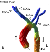
Fukui N, Toru KanahashiT, MatsubayashiJ, ImaiH, YoneyamaA, OtaniH, YamadaS, Takakuwa T. Morphogenesis of the pulmonary vein and left atrial appendage in human embryos and early fetuses. J Anatomy 2024, 244, 142-158, in press, https://doi.org/10.1111/joa.13941
Abstract
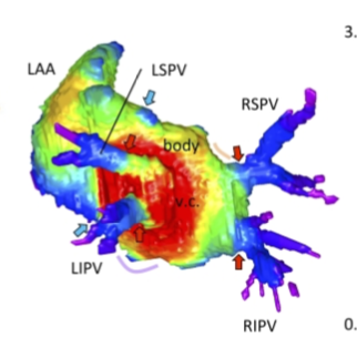
The left atrium wall has several origins, including the body, appendage, septum, atrial–ventricular canal, posterior wall, and venous component. Here, we describe the morphogenesis of left atrium based on high-resolution imaging (phase-contrast X-ray computed tomography and magnetic resonance imaging). Twenty-three human embryos and 19 fetuses were selected for this study. Three-dimensional cardiac images were reconstructed, and the pulmonary veins and left atrium, including the left atrial appendage, were evaluated morphologically and quantitatively. The positions of the pericardial reflections were used as landmarks for the border of the pericardial cavity. The common pulmonary vein was observed in three specimens at Carnegie stage 17–18. The pericardium was detected at the four pulmonary veins (left superior, left inferior, right superior, and right inferior pulmonary veins) at one specimen at Carnegie stage 18 and all larger specimens, except the four samples. Our results suggest that the position of the pericardial reflections was determined at two pulmonary veins (right and left pulmonary vein) and four pulmonary veins almost simultaneously when the dorsal mesocardial connection between the embryo and heart regressed. The magnetic resonance images and reconstructed heart cavity images confirmed that the left atrium folds were present at the junction between the body and venous component. Three-dimensional reconstruction showed that the four pulmonary veins entered the dorsal left atrium tangentially from the lateral to the medial direction. More specifically, the right pulmonary veins entered at a greater angle than the left pulmonary veins. The distance between the superior and inferior pulmonary veins was shorter than that between the left and right pulmonary veins. Three-dimensional reconstruction showed that the venous component increased proportionally with growth. No noticeable differences to discriminate between the right and left parts of the venous component emerged, while the junction between the venous component and body gradually became inconspicuous but was still recognizable by the end of the observed early fetal period. The left superior pulmonary vein had the smallest cross-sectional area and most flattened shape, whereas the other three were similar in area and shape. The left atrial appendage had a large volume in the center and extended to the periphery as a lobe-like structure. The left atrial appendage orifice increased in the area and tended to become flatter with growth. The whole left atrium volume^(1/3) increased almost proportionally with growth, parallel to the whole heart volume. This study provided a three-dimensional and quantitative description of the developmental process of left atrium, comprising the venous component and left atrial appendage formation, from the late embryonic to the early fetal stages.