


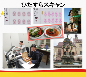
平日はひたすらスキャン
6/10−6/26、ドイツゲッチンゲン大学に共同研究者の山田先生、宮崎さん(山田研)、五十嵐さんの3名が、画像取得に出張いたしました。貴重なBlechshmidt collectionのうちの、連続組織標本をスキャニングしました。ご苦労さまです。
ゲッチンゲン大学解剖学教室は、昨年に引き続いての訪問になります。これからも定期的に伺うことになると思います。
|
||||
  |
平日はひたすらスキャン 6/10−6/26、ドイツゲッチンゲン大学に共同研究者の山田先生、宮崎さん(山田研)、五十嵐さんの3名が、画像取得に出張いたしました。貴重なBlechshmidt collectionのうちの、連続組織標本をスキャニングしました。ご苦労さまです。 ゲッチンゲン大学解剖学教室は、昨年に引き続いての訪問になります。これからも定期的に伺うことになると思います。 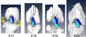 張、高石、樋口君の論文が Plos oneに受諾されました。 ラットの膝関節の発生を、EFICを用いて検討しました。特にACL, PCL靭帯の形成に着目しました。 樋口君の卒業論文を基にして、例数を増やしてまとめました。
14. Xiangkai Zhang; Tomoki Aoyama; Ryota Takaishi; Shinya Higuchi; Shigehito Yamada; Hiroshi Kuroki; Tetsuya Takakuwa, Spatial change of cruciate ligaments in rat embryo knee joint by three-dimensional reconstruction.PLoS One. 2015 Jun 22;10(6):e0131092. doi: 10.1371/journal.pone.0131092. eCollection 2015. AbstractThis study aimed to analyze the spatial developmental changes of rat cruciate ligaments by three-dimensional (3D) reconstruction using episcopic fluorescence image capture (EFIC). Cruciate ligaments of Wister rat embryos between embryonic day (E) 16 and E20 were analyzed. Samples were sectioned and visualized using EFIC. 3D reconstructions were generated using Amira software. The length of the cruciate ligaments, distances between attachment points to femur and tibia, angles of the cruciate ligaments and the cross angle of the cruciate ligaments were measured. The shape of cruciate ligaments was clearly visible at E17. The lengths of the anterior cruciate ligament (ACL) and posterior cruciate ligament (PCL) increased gradually from E17 to E19 and drastically at E20. Distances between attachment points to the femur and tibia gradually increased. The ACL angle and PCL angle gradually decreased. The cross angle of the cruciate ligaments changed in three planes. The primordium of the 3D structure of rat cruciate ligaments was constructed from the early stage, with the completion of the development of the structures occurring just before birth. |
|||