植田さんの卒業研究が、Anatomical Recに受諾されました。
ヒト胚子期の中腸の発生過程でみられる、腸の回転と臍帯内への生理的ヘルニアについての定量的な研究です。
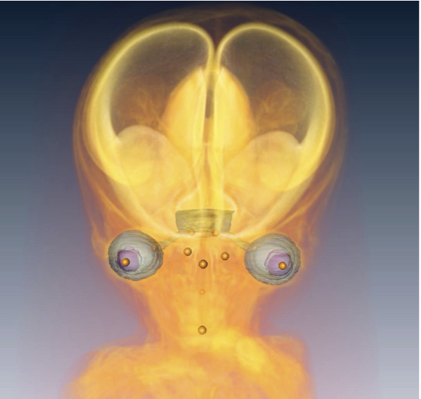
- CS14-23の中腸を3次元再構築し検討。
- CS16までに十二指腸、結腸直腸は正中から偏位
- CS17以降臍帯ヘルニア確認
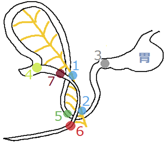
- CS18;ヘアピン状で、中腸間膜管動脈(SMA)がその直線部を並走、回盲部はSMAの左側
- CS19以降;小腸の急速な伸長、複雑な走行、回転はほぼ90度となり、尾側移動
- CS20まで;腸ループの立ち上がり部は右頭側から尾側へ移動、SMAに対して180度回転
- 腸ループの着地部はSMAの左尾側で著変なし
- 腸ループの動きは分化発生(differential growth)による受動的なものと考えられる
19. Ueda Y, Yamada S, Uwabe C, Kose K, Takakuwa T, Intestinal rotation and physiological umbilical herniation during the embryonic period, Anatomical Record 299, 197-206, 2016, DOI: 10.1002/ar.23296
ABSTRACT
Drastic changes occur during the formation of the intestinal loop (IL), including elongation, physiological umbilical herniation (PUH), and midgut rotation. Fifty-four sets of magnetic resonance images of embryos between Carnegie stage (CS) 14 and CS 23 were used to reconstruct embryonic digestive tract in three dimensions in the Amira program. Elongation, PUH, and rotation were quantified in relation to the proximal part of the superior mesenteric artery (SMA), designated as the origin. Up to CS 16, IL rotation was initially observed as a slight deviation of the duodenum and colorectum from the median plane. The PUH was noticeable after CS 17. At CS 18, the IL showed a hairpin-like structure, with the SMA running parallel to the straight part and the cecum located to the left. After CS 19, the IL began to form a complex structure as a result of the rapid growth of the small intestinal portion. By CS 20, the IL starting point had moved from the right cranial region to an area caudal to the origin, though elongation of the duodenum was not conspicuous—this was a change of almost 180° in position. The end of the IL remained in roughly the same place, to the left of and caudal to the origin. Notably, the IL rotated around the origin only during earlier stages and gradually moved away, running transversely after CS 19. The movements of the IL may be explained as the result of differential growth, suggesting that IL rotation is passive.