Osaka M, Ishikawa A, Yamada S, Uwabe C, Imai H, Matsuda T, Yoneyama A, Takeda T, Takakuwa T, Positional changes of the ocular organs during craniofacial development, Anatomical Record, 300(12), 2107–2114, 2017 DOI: 10.1002/ar.23588
ABSTRACT
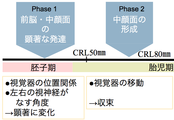
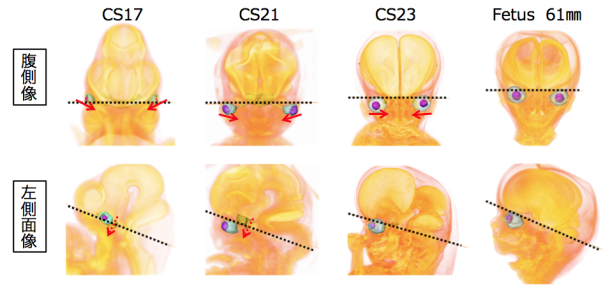
The present study aimed to describe the positional changes of the ocular organs during craniofacial development; moreover, we examined the relationships among the ocular organs and other internal structures. To do this, we traced the positions of the ocular organs in 56 human early fetal samples at different stages of development using high-resolution magnetic resonance imaging and phase-contrast X-ray computed tomography. The eyes were located on the lateral side in the ventral view at Carnegie stage (CS) 16, and then changed their positions medially during development. The eyes remained in the neurocranium until CS17. However, the eyes changed their positions medially and caudally in the viscerocranium after CS18. The positional relationship between the eyes and pituitary gland changed in the lateral view as development progressed. Specifically, they were close to each other at CS17, but moved apart during the later stages of development. These positional changes were also demonstrated quantitatively with morphometric analyses. Based on the present data, the positional changes of the eyes can be categorized into phases, as follows: Phase 1, dramatic positional changes (early fetal period until CS23); and Phase 2, mild positional changes (stabilized; early fetal period after CS23). Notably, all absolute lengths measured in the present study linearly increased as the crown-rump length increased irrespective of the phase, while features of the measured angles and ratios differentially changed in Phases 1 and 2. The present data may help improve our understanding of both the normal and abnormal development of the ocular organs and craniofacial area.