


大坂さんの修論の前半部がAnatomical Recordに掲載されました。
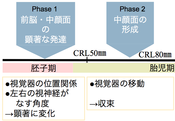
頭部・顔面形成にともなう眼の位置変化を胚子期から胎児期初期にかけて解析しました。
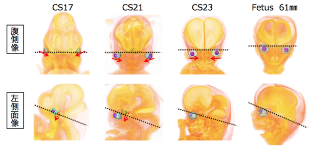
- CS16の正面像ではで眼球は、外側に位置し、発生がすすむとに内側に位置を変更
- CS17まで神経頭蓋内、CS18以降に内臓頭蓋の内側および尾側に位置を変更
- 眼球の位置変化が顕著な胚子期 (Phase1)と変化が少ない胎児期初期(Phase2)に大別され
- それらは Differential Growthの結果として説明できる
Osaka M, Ishikawa A, Yamada S, Uwabe C, Imai H, Matsuda T, Yoneyama A, Takeda T, Takakuwa T, Positional changes of the ocular organs during craniofacial development, Anatomical Record, 300(12), 2107–2114, 2017 DOI: 10.1002/ar.23588
ABSTRACT
The present study aimed to describe the positional changes of the ocular organs during craniofacial development; moreover, we examined the relationships among the ocular organs and other internal structures. To do this, we traced the positions of the ocular organs in 56 human early fetal samples at different stages of development using high-resolution magnetic resonance imaging and phase-contrast X-ray computed tomography. The eyes were located on the lateral side in the ventral view at Carnegie stage (CS) 16, and then changed their positions medially during development. The eyes remained in the neurocranium until CS17. However, the eyes changed their positions medially and caudally in the viscerocranium after CS18. The positional relationship between the eyes and pituitary gland changed in the lateral view as development progressed. Specifically, they were close to each other at CS17, but moved apart during the later stages of development. These positional changes were also demonstrated quantitatively with morphometric analyses. Based on the present data, the positional changes of the eyes can be categorized into phases, as follows: Phase 1, dramatic positional changes (early fetal period until CS23); and Phase 2, mild positional changes (stabilized; early fetal period after CS23). Notably, all absolute lengths measured in the present study linearly increased as the crown-rump length increased irrespective of the phase, while features of the measured angles and ratios differentially changed in Phases 1 and 2. The present data may help improve our understanding of both the normal and abnormal development of the ocular organs and craniofacial area.