Kitazawa H, Fujii S, Ishiyama H, Matsubayashi J, Ishikawa A, Yamada S, Takakuwa T. Nascent nephrons during human embryonic development: Spatial distribution and relationship with urinary collecting system. J Anatomy 2021; 238, 455-466, in press.DOI: 10.1111/JOA.13308
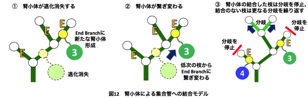
腎小体は尿路樹の末梢枝のみに結合するという観察を説明するモデル
Abstract
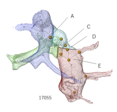
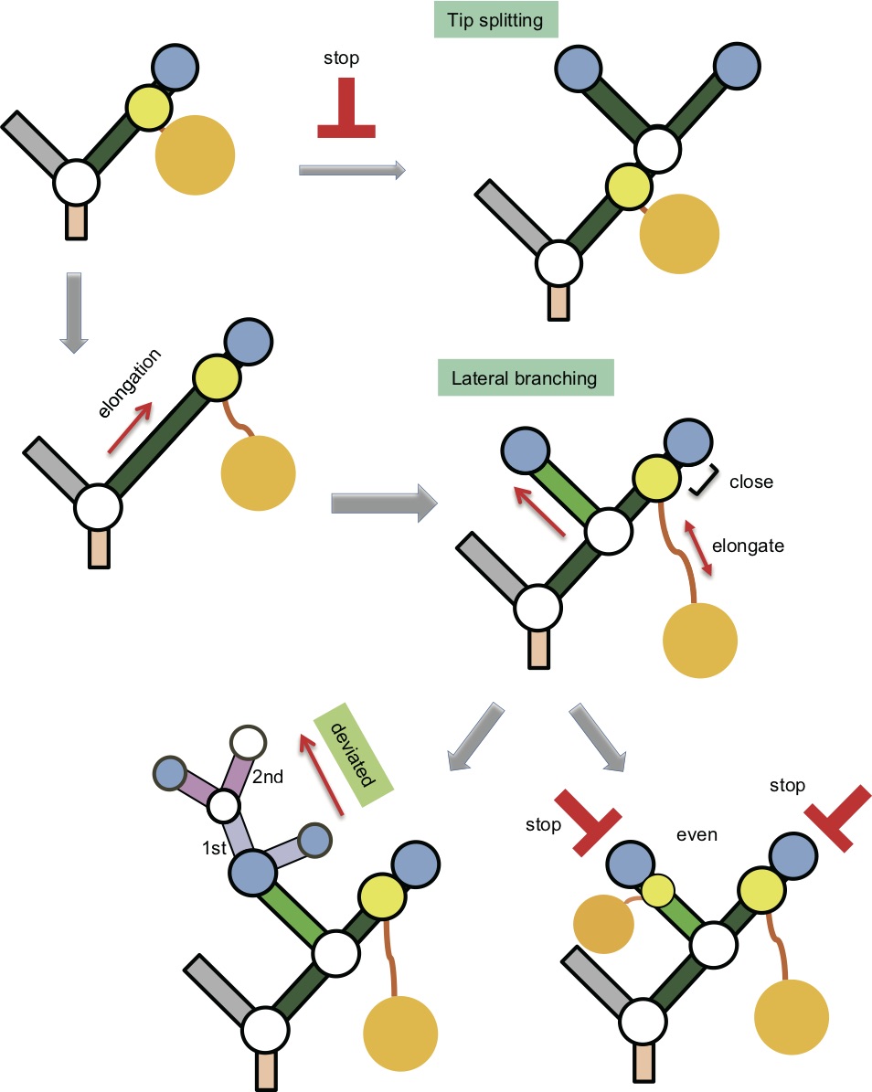
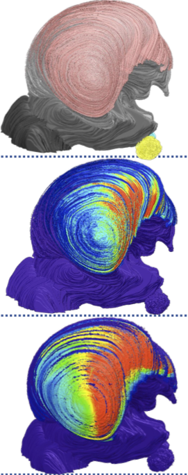
The two major components of the metanephros, the urinary collecting system (UCS) and nephron, have different developmental courses. Nephron numbers vary widely between species and individuals and are determined during fetal development. Furthermore, the development of nascent nephrons may contribute to the expansion of the proximal part of the UCS. This study investigated the distribution of nascent nephrons and their interrelationship with UCS branches during human embryogenesis. We obtained samples from 31 human embryos between Carnegie stages (CSs) 19 and 23 from the Kyoto Collection at the Congenital Anomaly Research Center of Kyoto University in Japan. Serial histological sections of the metanephros with the UCS were digitalized and computationally reconstructed for morphological and quantitative analyses. The three-dimensional (3D) coordinates for the positions of all UCS branch points, end points, attachment points to nascent nephrons (APs), and renal corpuscles (RCs) were recorded and related to the developmental phase. Phases were categorized from phase 1 to phase 5 according to the histological analysis. The UCS branching continued until RCs first appeared (at CS19). End branches with attached nascent nephrons (EB-AP[+]) were observed after CS19 during the fifth generation or higher during the embryonic period. The range of end branch and EB-AP(+) generation numbers was broad, and the number of RCs increased with the embryonic stage, reaching 273.8 ± 104.2 at CS23. The number of RCs connected to the UCS exceeded the number not connected to the UCS by CS23. The 3D reconstructions revealed RCs to be distributed around end branches, close to the surface of the metanephros. The RCs connected to the UCS were located away from the surface. The APs remained near the end point, whereas connecting ducts that become renal tubules were found to elongate with maturation of the RCs. Nascent nephrons in RC phases 3-5 were preferentially attached to the end branches at CS22 and CS23. The mean generation number of EB-AP(−) was higher than that of EB-AP(+) in 19 of 22 metanephros and was statistically significant for eight metanephros at CS22 and CS23. The ratio of the deviated branching pattern was almost constant (29%). The ratio of the even branching pattern with EB-AP(+) and EB-AP(+) to the total even branching pattern increased with CS (9.2% at CS21, 19.2% at CS22, and 45.4% at CS23). Our data suggest the following: EB-AP(+) may not branch further at the tip (i.e., by tip splitting), but branching beneath the AP (lateral branching) continues throughout the embryonic stages. Our study provides valuable data that can further the understanding of the interactions between the UCS and nascent nephrons during human embryogenesis.
Fujii S, Muranaka T, Matsubayashi J, Yamada S, Yoneyama A, Takakuwa T. Bronchial tree of the human embryo: categorization of the branching mode as monopodial and dipodial, PLoS One 2021, Published: January 15, 2021, https://doi.org/10.1371/journal.pone.0245558
Abstract
Some human organs are composed of bifurcated structures. Two simple branching modes—monopodial and dipodial—have been proposed. With monopodial branching, child branches extend from the sidewall of the parent branch. With dipodial branching, the tip of the bronchus bifurcates. However, the branching modes of the human bronchial tree have not been elucidated precisely. A total of 48 samples between Carnegie stage (CS) 15 and CS23 belonging to the Kyoto Collection were used to acquire imaging data with phase-contrast X-ray computed tomography. Bronchial trees of all samples were three-dimensionally reconstructed from the image data. We analyzed the lobar bronchus, segmental bronchus, and subsegmental bronchus. After calculating each bronchus length, we categorized the branching mode of the analyzed bronchi based on whether the parent bronchus was divided after generation of the analyzed bronchi. All lobar bronchi were formed with monopodial branching. Twenty-five bifurcations were analyzed to categorize the branching mode of the segmental and subsegmental bronchi; 22 bifurcations were categorized as monopodial branching, two bifurcations were not categorized as any branching pattern, and the only lingular bronchus that bifurcated from the left superior lobar bronchus was categorized as dipodial branching. The left superior lobar bronchus did not shorten during the period from CS17 or CS18, when the child branch was generated, to CS23. All analyzed bronchi that could be categorized, except for one, were categorized as monopodial branching. The branching modes of the lobar bronchus and segmental bronchus were similar in the mouse lung and human lung; however, the modes of the subsegmental bronchi were different. Furthermore, remodeling, such as shrinkage of the bronchus, was not observed during the analysis period. Our three-dimensional reconstructions allowed precise calculation of the bronchus length, thereby improving the knowledge of branching morphogenesis in the human embryonic lung.
51. Terashima, M., Ishikawa A., Männer J., Yamada S.&Takakuwa T. (2021) Early development of the cortical layers in the human brain. Journal of Anatomy, 239, 1039–1049. https://doi.org/10.1111/joa.13488
二次口蓋癒合に伴う口鼻腔領域の形態変化:形態測定学的手法を用いた三次元解析 野原 葵 Dynamic change in oronasal region during secondary palate fusion: Three-dimensional analysis using morphometrics
57. Nohara A, Owaki N, Matsubayashi J, Katsube M, Imai H, Yoneyama A, Yamada S, Kanahashi T, Takakuwa T. Morphometric analysis of secondary palate development in human embryos. J Anatomy, 2022, in press
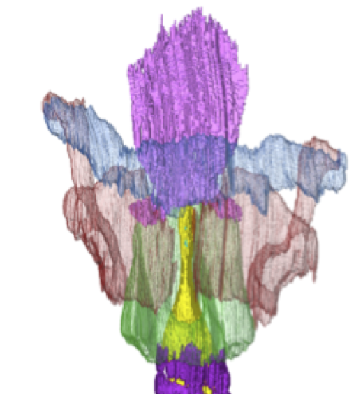
ヒト胚子期・胎児期初期における喉頭軟骨・気管軟骨の3次元的解析 山崎 優 Three-dimensional analysis of the human laryngeal and tracheal cartilages during the late embryonic and early fetus period
54. Yamazaki Y, Kanahashi T, Yamada S, Männer J, Takakuwa T. Three-dimensional analysis of human laryngeal and tracheobronchial cartilages during the late embryonic and early fetal period. Cells Tissues Organs, 2021 in press