


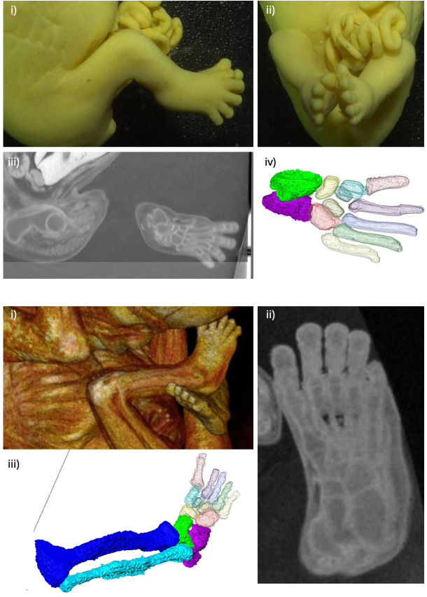
足関節骨格器系の形成についての論文が、Anat Recに受諾されました。胎児期の一時期、足関節が尖足、内反位を示す、生理的内反足を示します。この肢位の正しい理解は、内反足という先天性疾患の理解、診断の向上の面から重要です。
弊研究室で、5年ほど前から継続して解析している内容の初報になります。
- 生理的内反足の姿勢とその変化をより深く理解するため、胎生後期から胎児期初期の標本を高解像度CT, MRIを用いて撮像、骨格要素を3次元再構成。
- 前方、側方、後方、および足底の視点から、経時的な変化を形態学的および形態計測学的に分析
- 足首関節の底屈の減少は後足部の位置変化を著しく複雑化
- 後足部の持続的な回外、足軸に沿った前足部の回内、足首関節の底屈の減少は、生理的内反足の発達に重要な寄与
- 前足部と後足部の捻転と、脛骨距骨関節の傾斜に起因する足首関節の外転は顕著
- 「差別的成長」の概念が足首関節と足の形態形成に当てはまる可能性があることを示唆
74. Takakuwa T, Matsuda K, Yamato Y, Tamura S, Kimura K, Fujii S, Kanahashi T, Yoneyama A, Imai H, Otani H, Yamada S. Changes in the position of skeletal elements of the ankle and foot during late embryonic and fetal periods. Anat Rec (Hoboken) 2025, in press
The morphology of the ankle joint and foot during early development exhibits distinct differences from that observed in adults, with physiological clubfoot being a well-documented phenomenon. To better understand this posture and its transformation, the skeletal elements in this region were three-dimensionally reconstructed using high-resolution phase-contrast X-ray computed tomography and magnetic resonance imaging of specimens (n=23) during the late embryonic and early fetal periods, before joint cavity formation. Sequential changes were analyzed both morphologically and morphometrically from anterior, lateral, posterior, and plantar views. The reduction in plantar flexion of the ankle joint rendered the positional change of the hindfoot substantially more complex, and three-dimensional reconstruction facilitated its comprehension. Continuous supination of the hindfoot, pronation of the forefoot along the foot axis, and reduced plantar flexion of the ankle joint were identified as key postural changes that contributed to the development of temporal physiological clubfoot, initiated as early as the late embryonic period. Twisting between the forefoot and hindfoot and the abduction of the ankle joint, resulting from the obliquity of the tibia–talus joint, were substantial. The offset effect of the two angle changes conceals such changes in most previous studies. Changes in the shape of the tarsus bones, especially the calcaneus and talus, affected the mutual and adjacent bone positions, indicating that the concept of “differential growth” may apply to ankle-joint and foot morphogenesis. Findings of the present study are expected to enhance understanding of the pathogenesis and mechanisms underlying clubfoot and facilitate fetal diagnosis via morphological assessments.