


海外くんの卒業論文がAnatomical recordに掲載されました。卒業後、 Office assistantとして2年かけて仕上げてくれました。教科書には、胃の動きは”下降”、”頭尾軸に対する回転”、”背腹軸に対する回転”と3つの動きにわけて説明されています。しかしながら実際の動きは立体空間的でありそう単純ではありませんでした。表紙絵に採用されました。
8. Morphogenesis and three-dimensional movement of the stomach during the human embryonic period,
2014 May;297(5):791-7. doi: 10.1002/ar.22833.
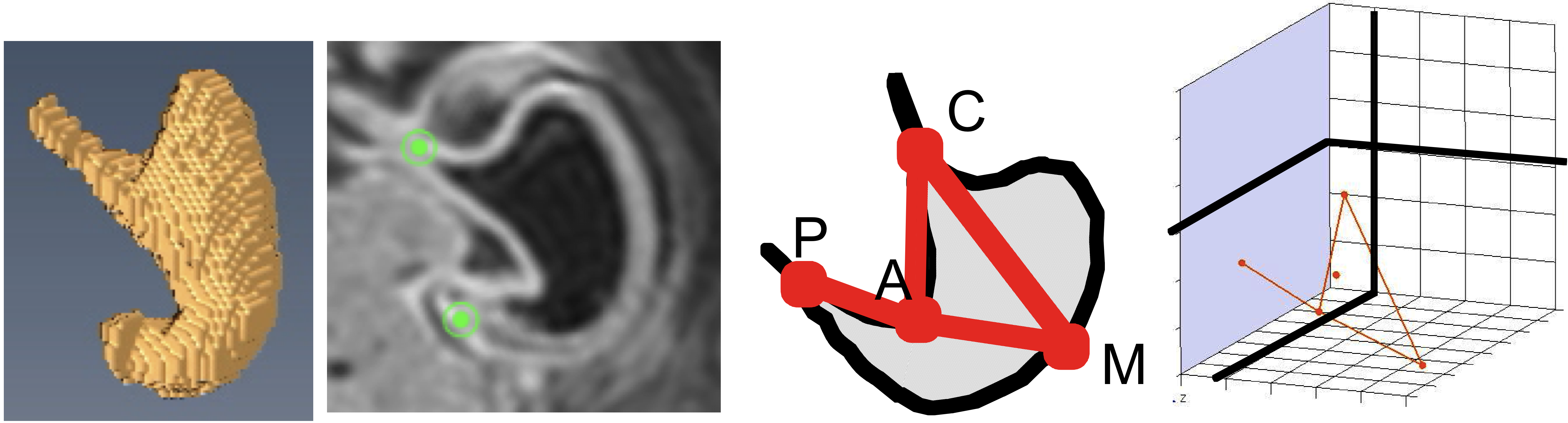
- 377例の胚子MR画像を用いて、CS16-23の胃の形態形成と動きを検討
- stageごとに特徴的な形態
- CS18; 胃角、胃底部の隆起
- CS18-20; 胃角は90度程度であったが、それ以降鋭角
- CS20; 噴門、幽門の分化がみられた。
- 大弯(M)の3次元的な動き(M), は噴門(C)、幽門(P)の動きと大きく異なる。
- C、PはCS16-23の間正中矢状面上にほぼ存在
- Mは尾側、左側にCS22まで大きく移動
- CPは左右軸を中心に回転
- 胃の最大平面CPMはおもに頭尾軸を中心に回転
- 胃の偏位とdifferential growthにより胃は左側、尾側に移動するように見えると推察
本研究の立体画像元データの一部はMorphoMuseuMに受諾されました。
20. Nako A, Kaigai N, Shiraki N, Yamada S, Uwabe C, Kose K, Takakuwa T, 3D models related to the publication: Morphogenesis of the stomach during the human embryonic period, MorphoMuseuM, in press
ABSTRACT
The stomach develops as the local widening of the foregut after Carnegie stage (CS) 13 that moves in a dramatic and dynamic manner during the embryonic period. Using the magnetic resonance images of 377 human embryos, we present the morphology, morphometry, and three-dimensional movement of the stomach during CS16 and CS23. The stomach morphology revealed stage-specific features. The angular incisura and the cardia were formed at CS18. The change in the angular incisura angle was approximately 90° during CS19 and CS20, and was <90° after CS 21. The prominent formations of the fundus and the pylorus differentiate at around CS20. Morphometry of the stomach revealed that the stomach gradually becomes “deflected” during development. The stomach may appear to move to the left laterally and caudally due to its deflection and differential growth. The track of the reference points in the stomach may reflect the visual three-dimensional movement. The movement of point M, representing the movement of the greater curvature, was different from that of points C (cardia) and P (pyloric antrum). The P and C were located just around the midsagittal plane in all the stages observed. Point M moved in the caudal-left lateral direction until CS22. Moreover, the vector CP does not rotate around the dorsoventral axis, as widely believed, but around the transverse axis. The plane CPM rotated mainly around the longitudinal axis. The data obtained will be useful for prenatal diagnosis in the near future.