


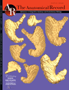
海外君、名古さんの論文で作成した胃の立体像が解剖学雑誌Anatomical Record 297巻5号の表紙に採用されるとともに、ビデオを結果として示すことのできるAR WOW-Video Articleの第1号の論文として採用されました。
N. Kaigai, A. Nako, S. Yamada, C. Uwabe, K. Kose and T. Takakuwa
Anat RecはAmerican Association of Anatomists (AAA)の公認雑誌で、100年以上の発行歴のある由緒ある雑誌です。永く世界の医学、解剖学、発生学の分野を牽引してきました。本雑誌は、さらに、最近のe-Page技術を取り入れたWOWという雑誌を本号から採用しました。これは、ビデオが論文の結果として重要な役割を果たす際にそのビデオが永久に保存できるようにしたものです。その第1号の論文として、海外君の胃の形態形成と動きについての論文が選ばれました。
Editorialでは、”Human Embryos are Turning on the e-Pages of AR”(ヒト胚子がARのe-Pageに灯をともす…)という題で, インタビューを受け、私たちの予想以上に讃えていただきました。(Anat Rec 297(5) ; 799–801, 2014)
Our rationale for imaging the human stomach during development.
“All of the authors are well-versed with the fact that the stomach develops as the local widening of the foregut at Carnegie Stage (CS) 13, as well as the morphology and position of the stomach in adults. But what are the developmental dynamics from the former to the latter? While I (Dr. Takakuwa) was a university student, I read a textbook that explained that the developmental dynamics of the stomach follow the order of linear movement along the caudal direction, rotation around the longitudinal (Z) axis, and rotation around the dorsoventral (X) axis. This explanation aroused my curiosity with regard to the position of the abdominal organs around the stomach, such as the esophagus, pancreas, and duodenum, which are restricted in their positions after CS17. For example, around CS20, movement of the stomach is restricted at both its entrance (cardia) and the exit (pyloric antrum) near the mid-sagittal plane.
We designed our study to sort out the dynamic process that places the stomach in its definitive position in the abdomen. Accordingly, we analyzed the external morphology and morphometry of the human embryonic stomach, as well as documented its precise 3D movements, using magnetic resonance (MR) imaging data of human embryos in the “Kyoto Collection”. We discovered that the line connecting the cardia and the pyloric antrum of the stomach does not rotate around the dorsoventral (X) axis, as widely believed, but rotates around the transverse (Y) axis. The stomach “appears” to move towards the left, laterally and caudally, as deflection and differential growth progresses. We found that the developmental morphology of the three-dimensionally reconstructed stomach was not “analogous” to that of adults or as described in recent textbooks. Rather, we found that the stomach’s developmental morphology is as documented in a study a century before (Lewis 1912), in which the stomach was precisely hand drawn by a special artist [note added by Editor: Lewis studied the stomachs of five human embryos that were 10 mm and 45 mm in length; Harvard Embryological Collection, Series 1000]. We are gratified that our MR imaging data of embryos enhance the value of the Kyoto Collection, not only as archives of historical specimens but also as useful research resources for the future.”
Contributed by Kaigai and colleagues, Kyoto University, Graduate School of Medicine.